
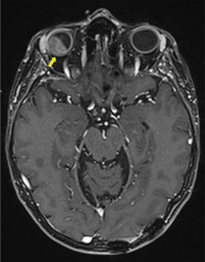
Figure 1
Contrast enhanced Axial T1 Dixon MRI-scan showing intra-orbital metastasis (arrow) invading all layers of the eyeball and the intra-conal fat.
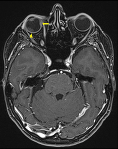
Figure 2
Contrast enhanced Axial T1 Dixon MRI-scan showing a second intra-orbital metastasis (arrow) invading all layers of the eyeball including sclera and a secondary retinal detachment (arrowhead).
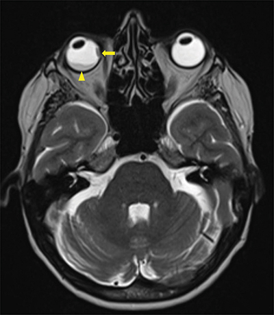
Figure 3
Axial T2 Dixon MRI-scan showing a second intra-orbital metastasis (arrow) invading all layers of the eyeball including sclera and a secondary retinal detachment (arrowhead).