
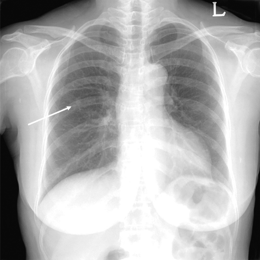
Figure 1
Initial chest radiograph shows an incidental nodule in the right upper lung field (arrow).
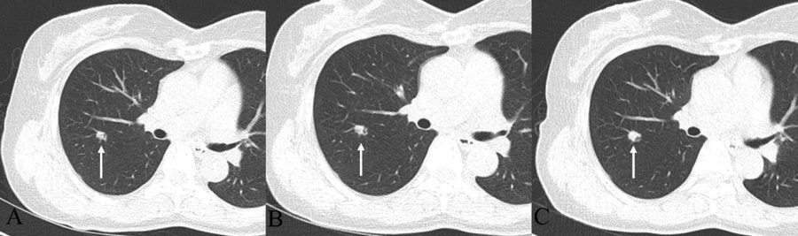
Figure 2
Chest CT with follow up. (A) 9-mm lobulated nodule with an internal air-bronchogram in the right upper lobe. (B) After 1 year, the nodule shows a minimal increase in size, 9 to 10 mm. (C) After 2 years later from the initial chest CT, the nodule changes from 10 to 13 mm in size, and the somewhat dilated internal bronchus are detected.
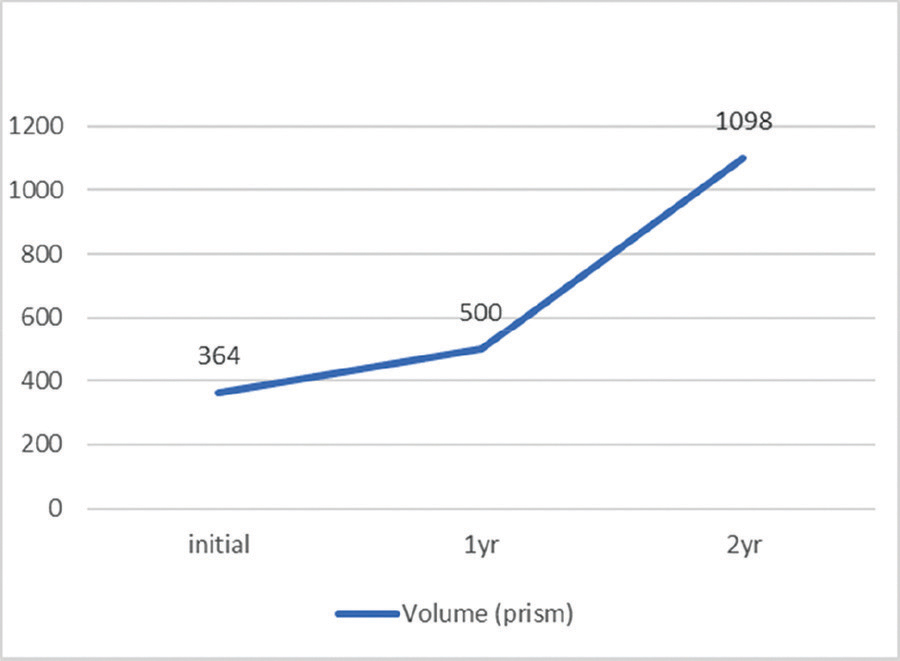
Figure 3
Volume: After 1 year, the volume doubling time from the initial state was 844.28 days, and the overall average volume doubling time after 2 years was 369.45 days.

Figure 4
Histopathology. (A–B) Hematoxylin-eosin staining slide images show that the papillary tumor is constituted of pseudostratified columnar epithelium covering the fibrotic core. Surrounding alveolar spaces are filled with mucus on frozen (A, ×40) and permanent sections (B, ×100). (B) The lesion shows multiple papillary fronds lined by stratified and pseudostratified columnar cells. Moderate stromal lymphocytic infiltration and psammoma bodies are noted in the focal peripheral area. (C) In partial areas, small papillae with oncocytic change within the adjacent alveolar spaces are mimicking to the histologic features of the spread through the air spaces of pulmonary adenocarcinoma (×100). (D) Most of the lesion is composed of ciliated or non-ciliated columnar, goblet, and cuboidal cells. There are neither architectural atypia nor cytological atypia (×100).