
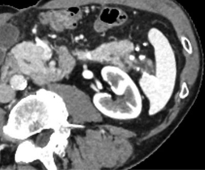
Figure 1
CT scan in arterial phase, showing caudal division of the tail of the pancreas and chronic pancreatitis changes posteriorly.
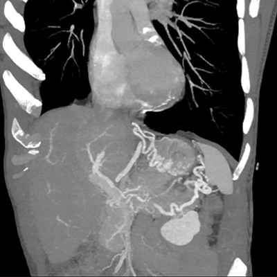
Figure 2
CT performed during portal phase, Maximum Intensity Projection 3D-reconstruction showing splenic vein thrombosis with subsequent gastro-epiploic and fundus varices.
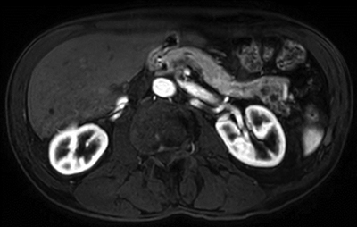
Figure 3
Axial cut in T1 DIXON sequence in arterial phase showing caudal division of the tail of the pancreas and chronic pancreatitis changes.
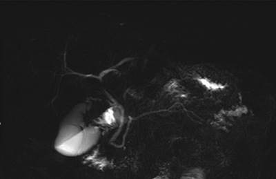
Figure 4
MR cholangiopancreatography, RARE sequence showing caudal division of the Wirsung duct and chronic pancreatitis changes.