
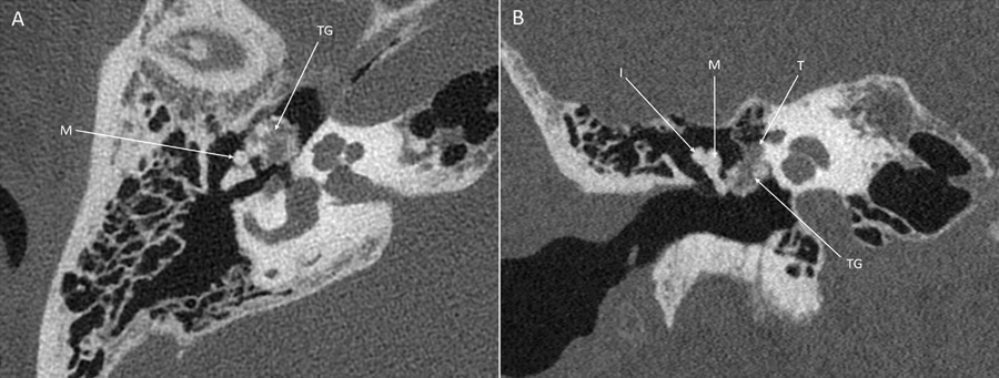
Figure 1
PCCT images (SIEMENS NAEOTOM Alpha) of the right temporal bone. (A) The axial image shows contact of the tophaceous gout (TG) with the malleus (M). (B) The coronal image shows contact with the tympanic segment of the facial nerve (T) and the malleus (M). The incus (I) is not affected.
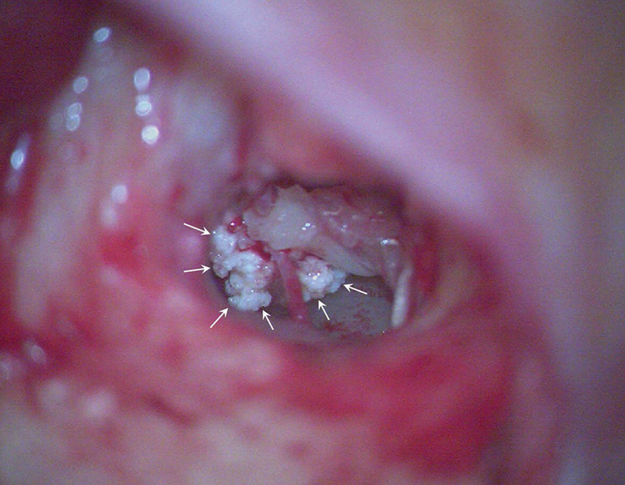
Figure 2
Intraoperative view of the middle ear, where the tophaceous gout is seen as a white and crystalline mass (arrows).
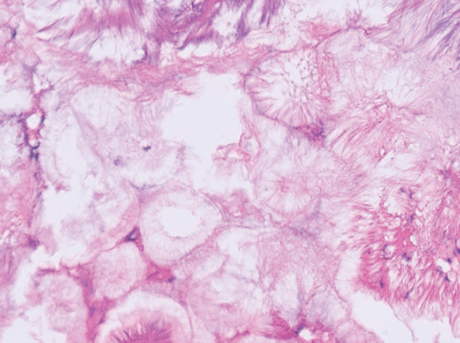
Figure 3
A hematoxylin and eosin-stained slide (scale 50 µm) shows the presence of empty needle-like structures with a feathery appearance, characteristic of gout tophi (Courtesy of A. Vanstapel MD, PhD).