
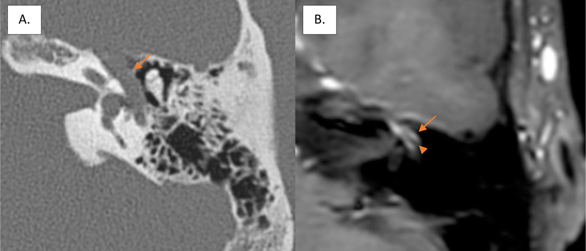
Figure 1
Enlargement of the tympanic segment of the fallopian canal (arrow) on axial CT scan of the left temporal bone (A) and intense contrast-enhancement (arrow) surrounding the facial nerve (arrowhead) on axial gadolinium-enhanced T1-weighted imaging (B).
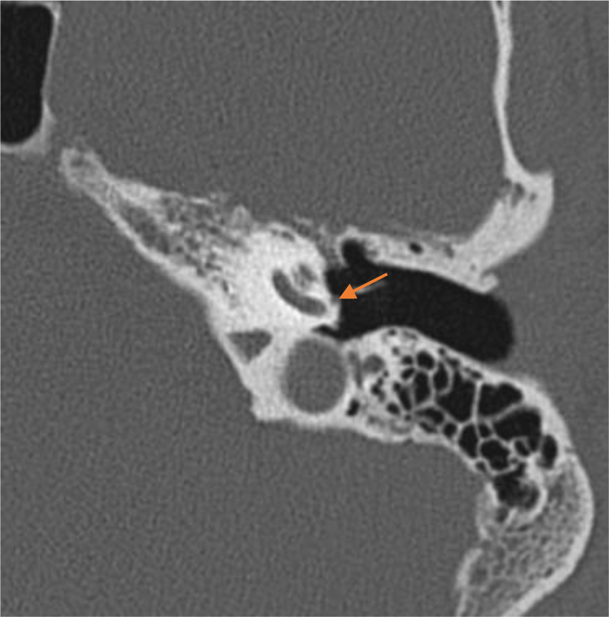
Figure 2
Axial CT scan showing the PSA on the cochlear promontory.
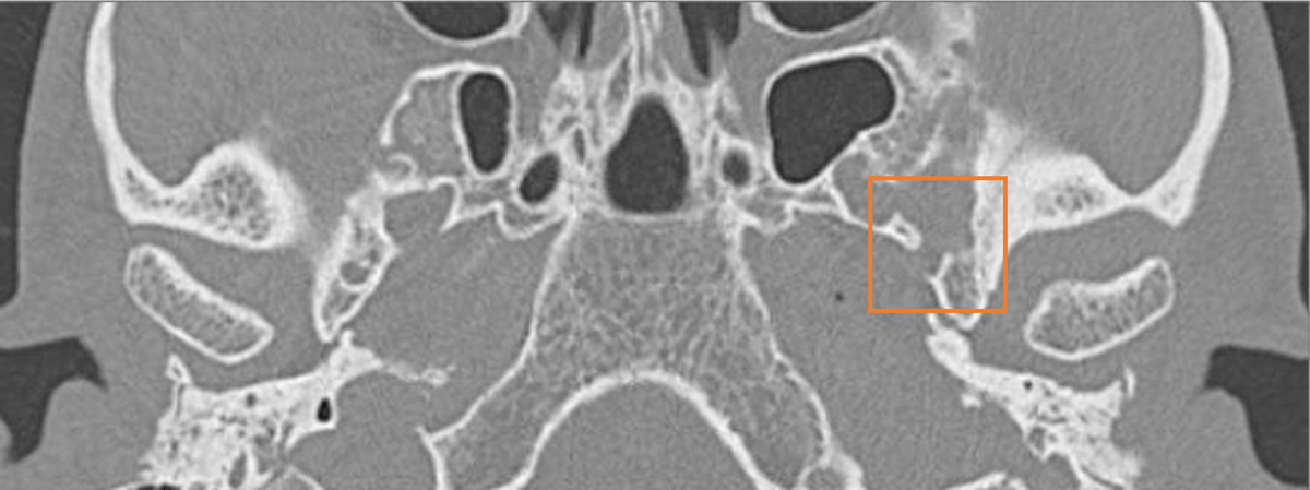
Figure 3
Axial CT scan showing the absence of the left foramen spinosum.