
Table 1
Patient History.
| PATIENT | AGE (y) | SEX | AFFECTED MUSCLE | VACCINATION |
|---|---|---|---|---|
| 1 | 74 | Male | Right brachialis muscle | Influenza |
| 2 | 66 | Male | Left brachioradialis muscle | COVID-19 |
| 3 | 64 | Male | Right deltoid muscle | COVID-19 |
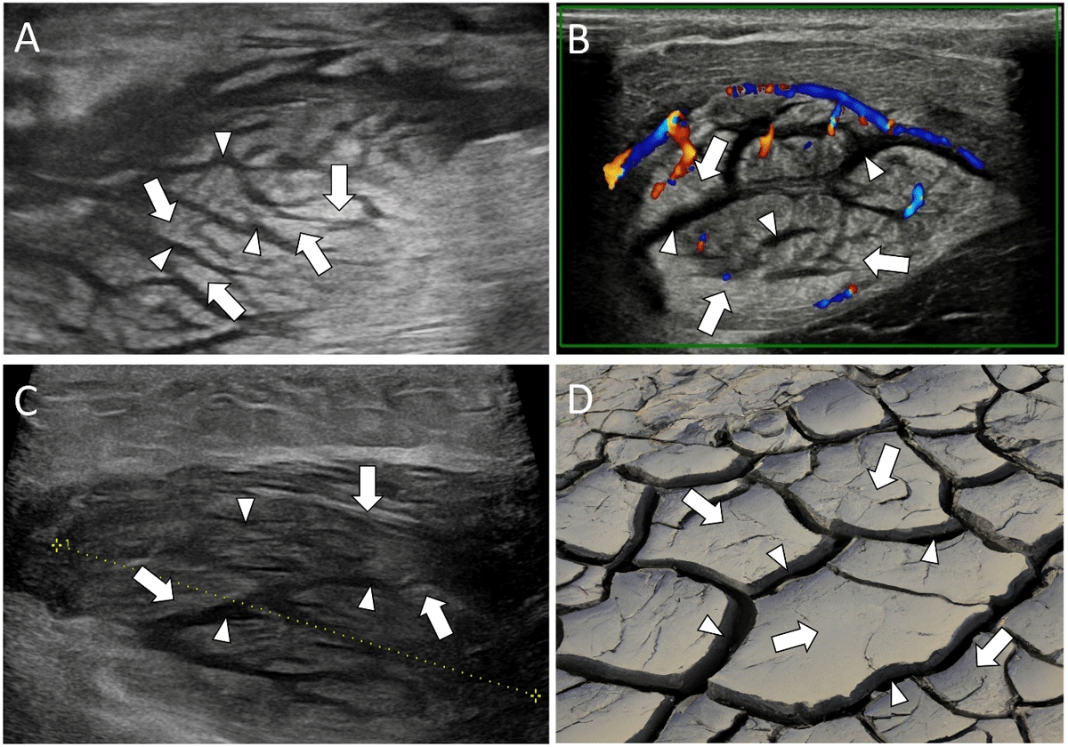
Figure 1
US showing a ‘cracked dry mud’ pattern of PM with swollen hyperechoic muscle fibers (white arrows) interspersed with hypoechoic bands (white arrowheads). A: Transverse image of the right brachialis muscle of patient 1. B: Transverse color Doppler image of the left brachioradialis muscle of patient 2 showing hypervascularity. C: Longitudinal image of the right deltoid muscle of patient 3. D: ‘Cracked dry mud’.
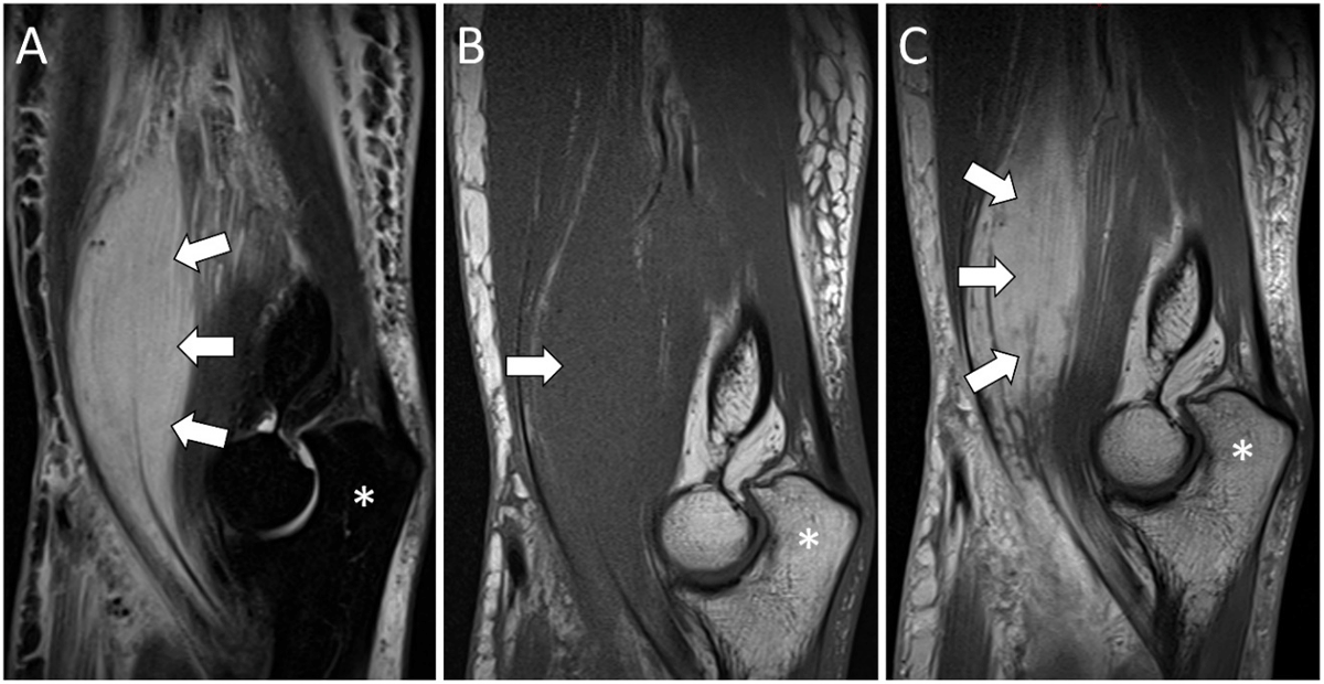
Figure 2
Sagittal MRI images of PM (white arrows) in the right brachialis muscle: hyperintense on STIR images (A), isointense compared to the muscle on T1WI (B), and a marked enhancement on post-contrast T1WI (C). Olecranon (*).
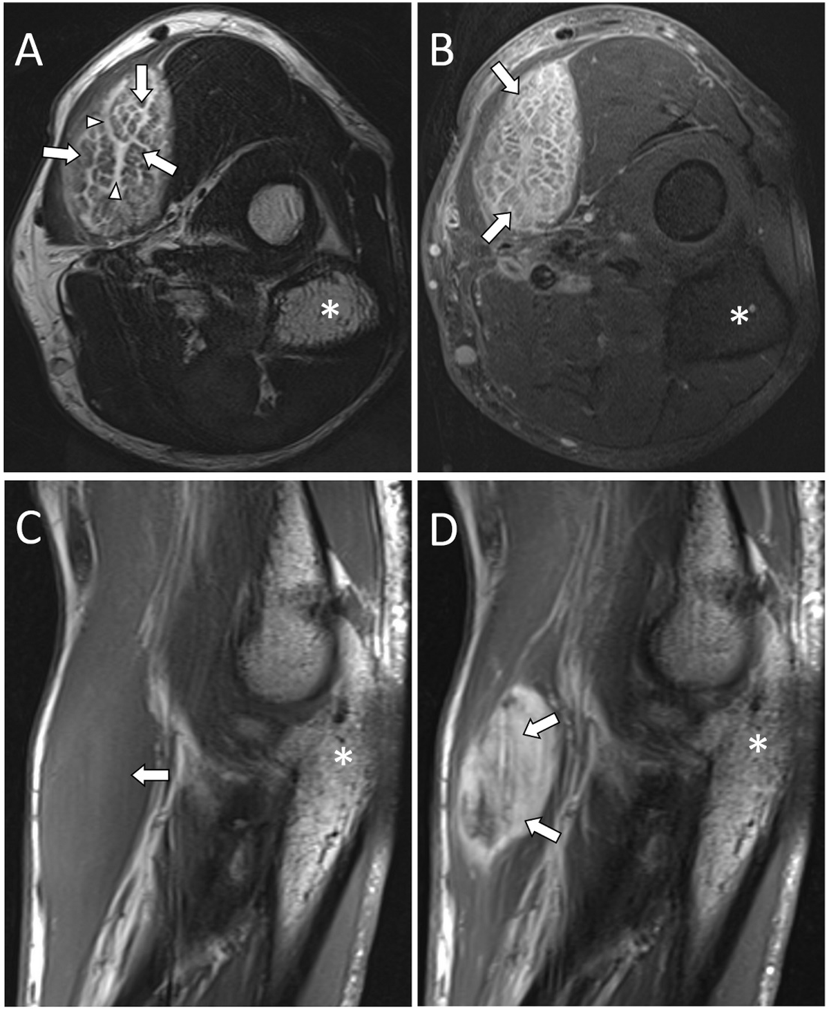
Figure 3
MRI images of PM in the left brachioradialis muscle. Axial T2WI (A), axial post-contrast T1WI fat-saturated (B), sagittal T1WI (C), and sagittal post-contrast T1WI (D) images demonstrate a T2 hyperintense lesion with a marked enhancement. The T2WI (A) shows hyperintense bands (white arrowheads) interspersed with intact muscle fibers (white arrows). Olecranon (*).
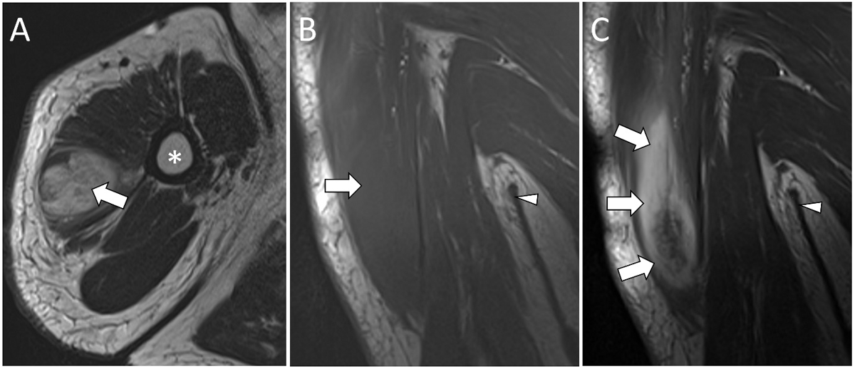
Figure 4
MRI images of PM in the right deltoid muscle demonstrate a hyperintense lesion (white arrow) on axial T2WI (A) and an isointense lesion on coronal T1WI (B) with a marked enhancement on coronal post-contrast T1WI (C) (white arrows). Humerus (*). Axilla (white arrowhead).