
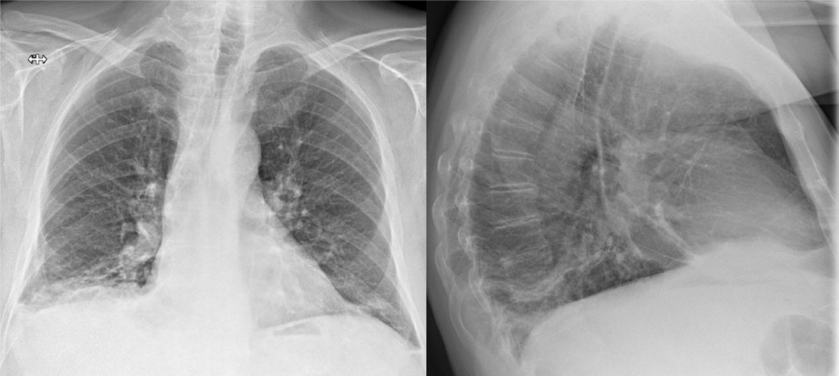
Figure 1
CXR with increased pleural fluid at the right lung basis and several dense strands.

Figure 2
HRCT with a ground-glass opacity surrounded by a crescentic consolidated zone (RHS) in the subpleural space of the RLL.

Figure 3
CT angiography with pathognomonic arterial filling defects.