
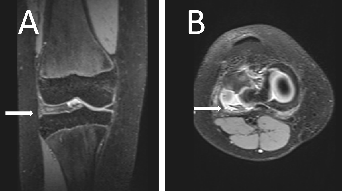
Figure 1
Proton density fat-saturated image of the girl’s knee showing a lateral discoid meniscus (arrow) in a coronal plane (A) and a degenerative, disinserted, meniscus with an anterior subluxation and posterior residual meniscal fragment facing the popliteal hiatus (arrow) in an axial plane (B).
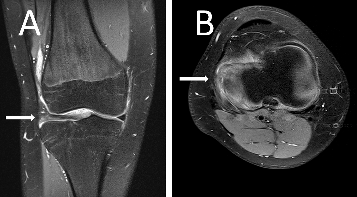
Figure 2
Proton density fat-saturated image of the boy’s knee showing a lateral discoid meniscus (arrow) in a coronal plane (A) and a complex posterior horn lesion (arrow) in of the meniscus in an axial plane (B).