

Figure 1
Intrasellar and suprasellar lesion in a 67-year-old female patient, which is hyperintense in on coronal T2WI (A), hypointense on coronal T1WI (B), and no enhancement is observed on contrast-enhanced T1WI (C) diagnosed as Rathke’s cleft cyst pathologically.
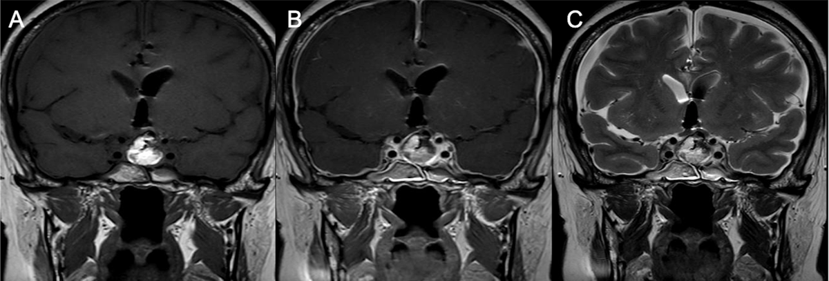
Figure 2
MRI after first surgery: Coronal T1WI (A), Coronal post-contrast T1WI (B) and coronal T2WI revealed signs of intracranial hypotension: right and left subdural collections and pachymeningeal thickening and enhancement.
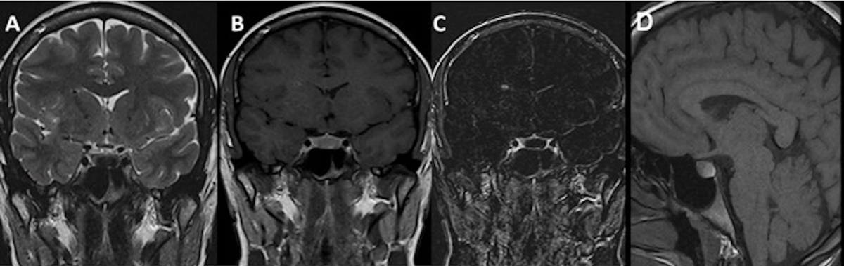
Figure 3
Intrasellar lesion in a 22-year-old female patient with a hypointense component on coronal T2WI (A), which is hyperintense on coronal (B) and sagittal (D) T1WI, and without enhancement on coronal contrast-enhanced substracted T1WI (C) diagnosed as Rathke’s cleft cyst pathologically.

Figure 4
Coronal T2WI (A), coronal T1WI (B), coronal contrast-enhanced T1WI (C) and axial FLAIR (D) images in a 73-year-old female patient show cystic intrasellar and suprasellar lesion with a non-enhancing intracystic nodule hyperintense to surrounding fluid on T1 and hypointense on T2.
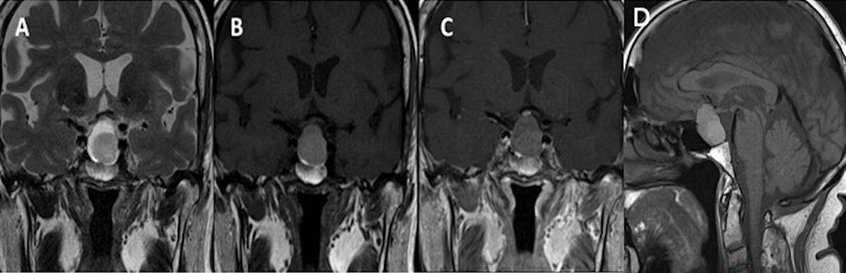
Figure 5
Ten-year follow-up of the same patient: Coronal T2WI (A), T1WI (B), post-contrast T1WI (C) and sagittal T1WI (D) show an increase in size of the lesion as well as the intracystic nodule.
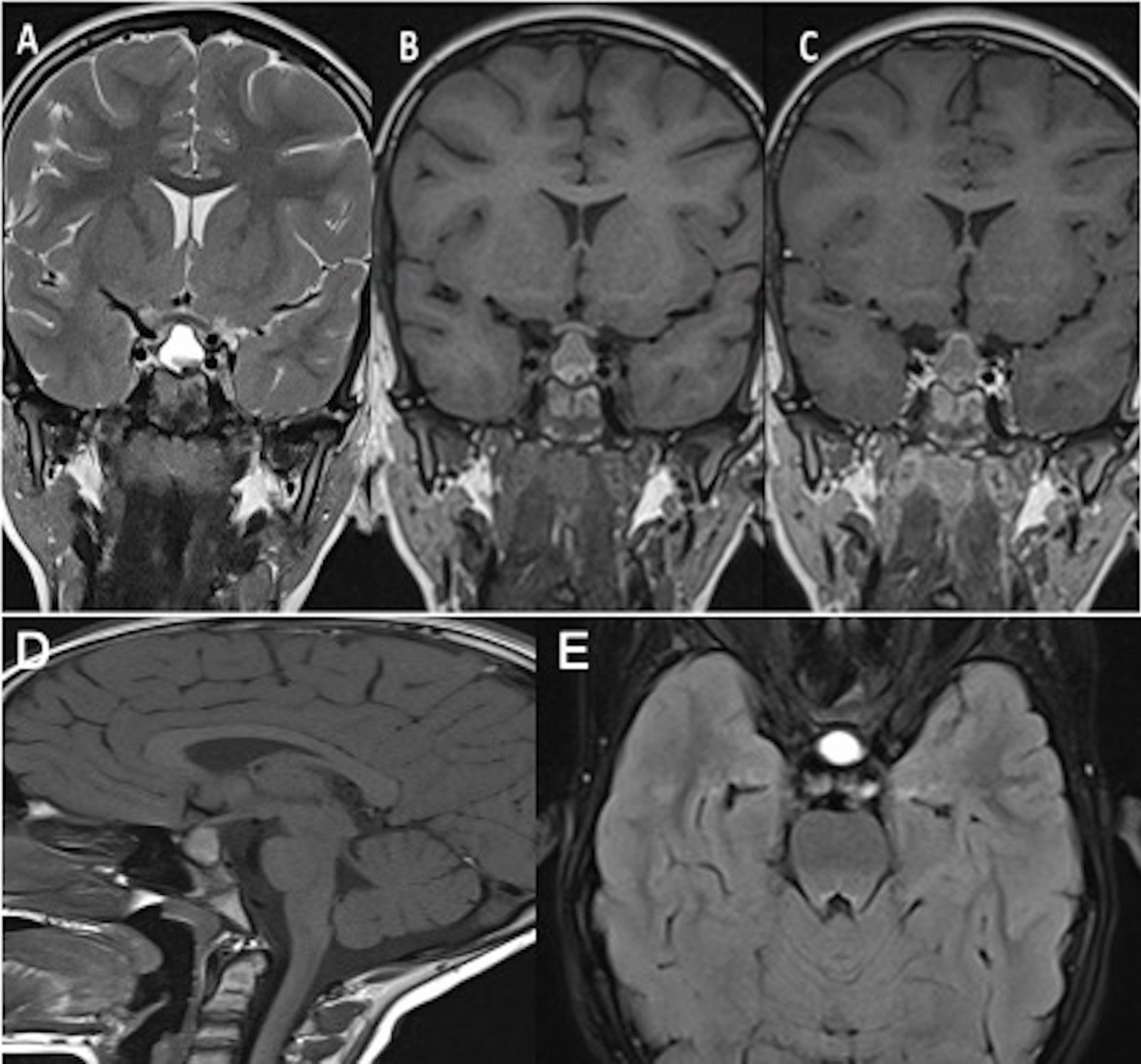
Figure 6
Coronal T2WI (A), T1WI (B), post-contrast T1WI (C), sagittal T1WI (D) and axial FLAIR (E) images demonstrate a sellar and suprasellar lesion with heterogeneous content in the lower part of the lesion and peripheric enhancement.