
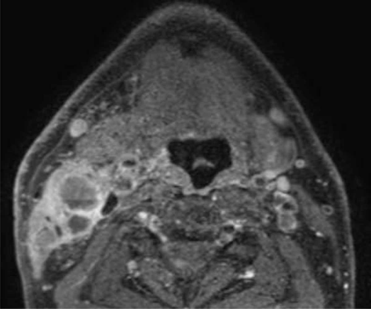
Figure 1
Patient with HPV-related oropharyngeal squamous cell carcinoma (not shown). Axial fat suppressed gadolinium enhanced MR section at the level of the tongue base shows predominantly cystic lymphadenopathy in the right neck (Level II).
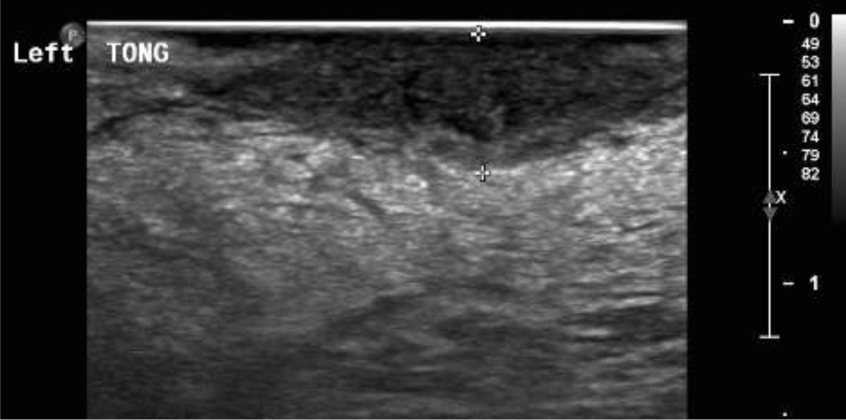
Figure 2
Patient with squamous cell carcinoma of the tongue. Intraoral ultrasound image showing the hypoechoic tongue tumor infiltrating the normal (echogenic) tongue musculature. The distance between the two calipers indicates the depth of invasion (DOI).