
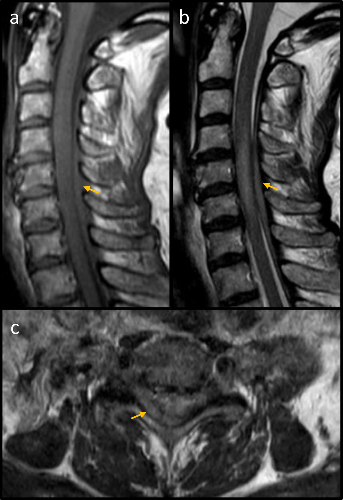
Figure 1
MRI of the cervical spine. (a) Sagittal T1 spin echo demonstrating an iso-intense space-occupying lesion (yellow arrow) restricted to the posterior epidural space. (b) Sagittal T2 spin echo showing a slightly hyper-intense mass (yellow arrow) in the posterior epidural space with corresponding myelopathy. (c) Axial T2 spin echo at vertebral level C6 showing a predominantly right-sided mass in the posterior epidural space (yellow arrow).
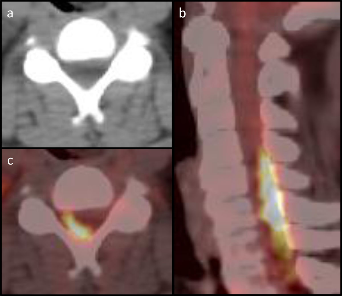
Figure 2
PET-CT of the cervical spine. (a) Axial CT at vertebral level C4 with barely visible posterior epidural mass. (b) Axial PET-CT of vertebral level C4 with notable FDG-avid disease in the spinal canal. (c) Sagittal PET-CT with FDG-avid disease posteriorly within the spinal canal.