
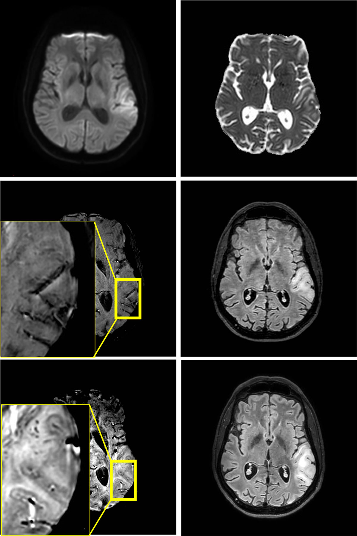
Figure 1
Edema in the left temporoparietal operculum without reduced diffusivity on MRI at 3T at first presentation (4 upper images). No microhemorrhages are found on the first scan, but they are present in juxtacortical location at one-week follow-up (2 lower images).
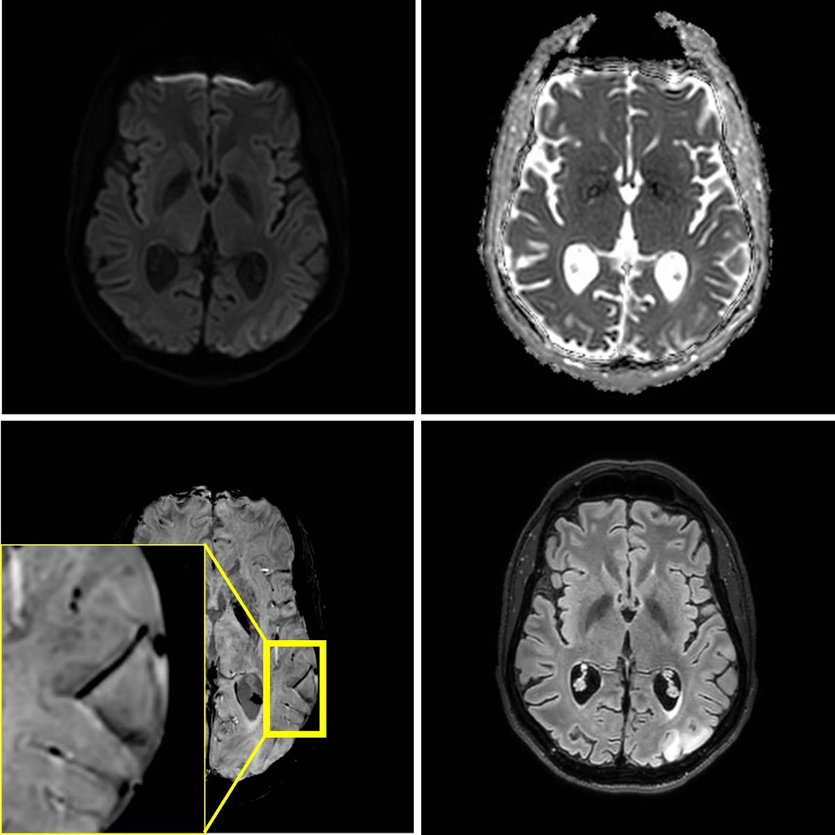
Figure 2
MRI at 3T performed three weeks later, because of a second stroke-like event. A similar lesion in the gyri of the left occipital lobe is present. Signal abnormalities in the previously affected area nearly normalized, but microhemorrhages persisted.
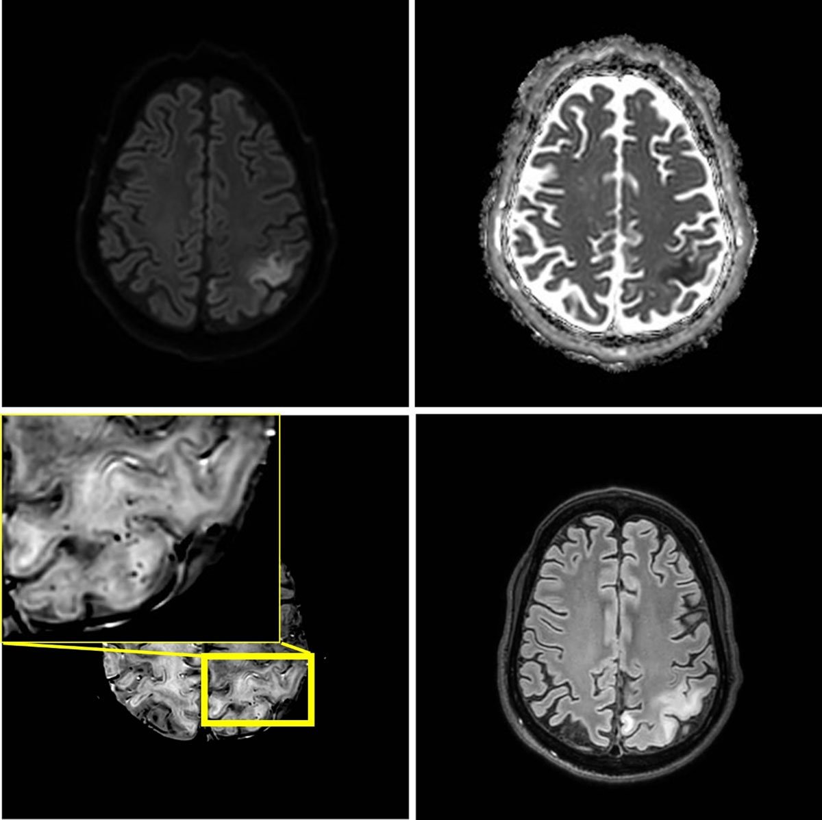
Figure 3
One month later, seven weeks after the initial episode, a third stroke-like event occurred. MRI shows a new area of edema in the left parietal lobe including the postcentral gyrus. The new lesion shows diffusion restriction and juxtacortical microhemorrhages.