
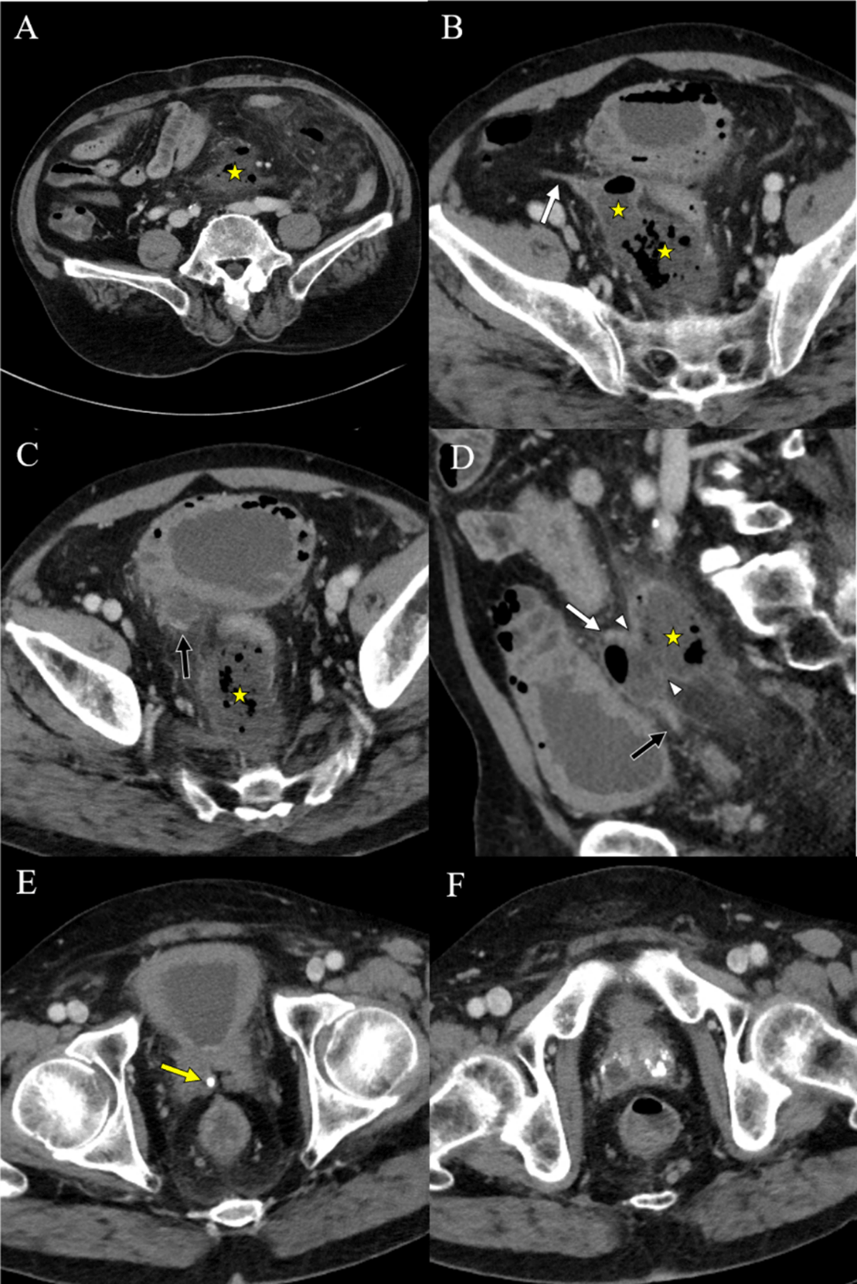
Figure 1
Pre-treatment CT images of a 65-year-old man with rupture of a vas deferens (VD) abscess. Axial and sagittal images (A–D) show a large abscess along the sigmoid mesocolon. Axial images (B, C) show the proximal and distal portion of right VD. A sagittal image (D) shows a wall defect of the VD adjacent to the abscess. An axial image (E) shows a tiny stone at the point where the VD joins the seminal vesicle. Axial images (F) show multifocal abscesses and dystrophic calcifications in the prostate. (asterisk: abscess, white arrow: proximal VD, black arrow: distal VD, arrowhead: VD wall defect, yellow arrow: VD stone).
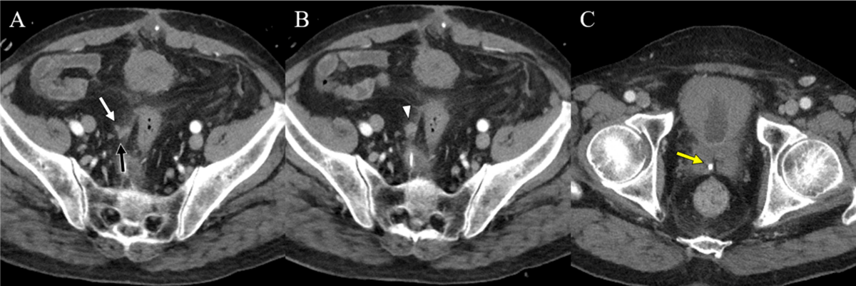
Figure 2
Follow-up CT images were taken after 11days. Axial images (A, B) show no abscess other than enhancing soft tissue (arrowhead) around the defect site of the VD wall. An axial image (C) shows a remnant stone at the distal VD. (white arrow: proximal VD, black arrow: distal VD, yellow arrow: VD stone).