
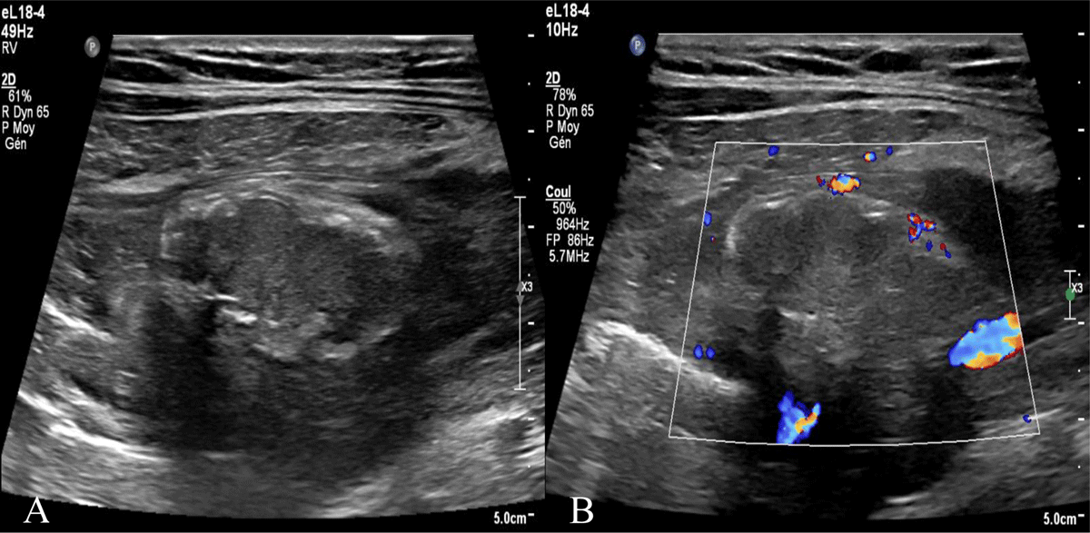
Figure 1
Right iliac fossa ultrasound. A) Superficial intraperitoneal oblong mass in the right iliac fossa of 35mm non-compressible, hyperechoic, surrounded by a subtle hypoechoic line. B) without internal vascularity.
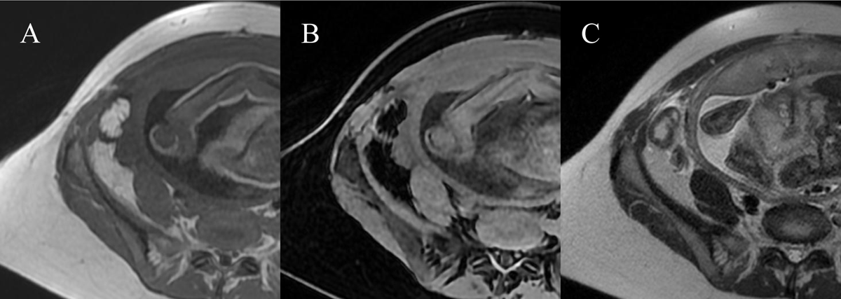
Figure 2
Right iliac fossa MRI. A) T1 sequence showed a anterior superficial intraperitoneal oblong mass in the right iliac fossa of 35mm hypersignal slightly less intense than normal fat with a thin peripheral hyposignal. B) T1FS sequence showed hyposignal of the mass with a fine periperal hypersignal. C) Hypersignal T2 of the mass and hypersignal T2 in the fat around.