
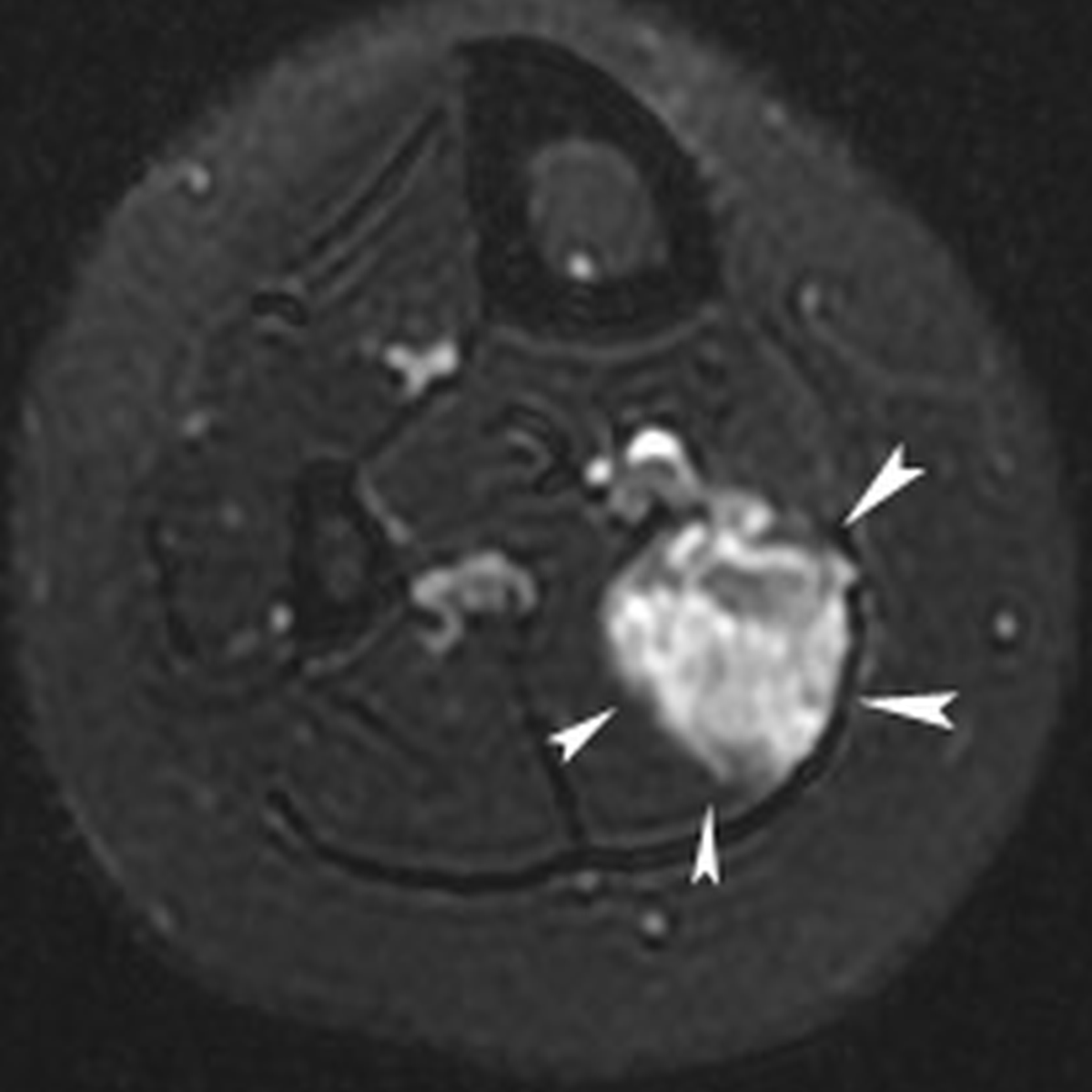
Figure 1a
Twenty-year-old female with chronic calf pain. T2-weigthed axial magnetic resonance image of the calf demonstrating an hyperintense mass lesion with a maximal diameter of 3.6 cm (white arrowheads) in the right soleus muscle, suggestive for low-flow vascular malformation.
Table 1
Patients demographics, clinical and radiological presentation of the VM.
| D/C/R PARAMETER | PATIENT 1 | PATIENT 2 | PATIENT 3 | PATIENT 4 |
|---|---|---|---|---|
| Age | 11 y | 14 y | 20 y | 19 y |
| Sex | female | female | female | female |
| Location of VM: Lower extremity | ||||
| upper leg | – | – | – | + |
| lower leg | – | + | + | – |
| Foot | + | – | – | – |
| Intramuscular | – | + | + | + |
| (lateral gastrocnemius) (soleus) (vastus intermedius) | ||||
| Subcutaneous tissue | + | – | – | – |
| Maximum diameter of the VM | 2 cm | 1, 8 cm | 3, 6 cm | 4, 3 cm |
| Patients’ symptoms: | ||||
| Local pain | + | + | + | + |
| Local swelling | + | + | + | + |
| Functional disability | + | + | - | + |
| Duration of symptoms since onset | NM | 4 y | 3 y | 3 y |
| Previous treatment | ||||
| sclerotherapy | – | + | + | – |
| . ethanol 96% | – | + | – | – |
| . sotradecol 3% | – | + | + (2x) | – |
| Interval between last sclerotherapy and RFA | ||||
| – | 8 months | 8 months | – | |
[i] VM = venous malformations.
RFA = radiofrequency ablation.
NM = not mentioned in electronic medical record.
Table 2
Patients’ clinical short- and long-term follow-up.
| PARAMETER | PATIENT 1 | PATIENT 2 | PATIENT 3 | PATIENT 4 |
|---|---|---|---|---|
| Short & midterm FU (1 year) | ||||
| Local pain | – | – | – | – |
| Local swelling | – | – | – | – |
| Functional disability | – | – | + | – |
| Procedure-related complication | – | – | + | – |
| Long-term follow-up (until 1/4/2021) | ||||
| FU interval (years) | 10 y | 11 y | 7 y | 6 y |
| Local pain | – | – | – | – |
| Local swelling | – | – | – | – |
| Functional disability | – | – | –/+ | – |
| Procedure-related complication | – | – | + | – |
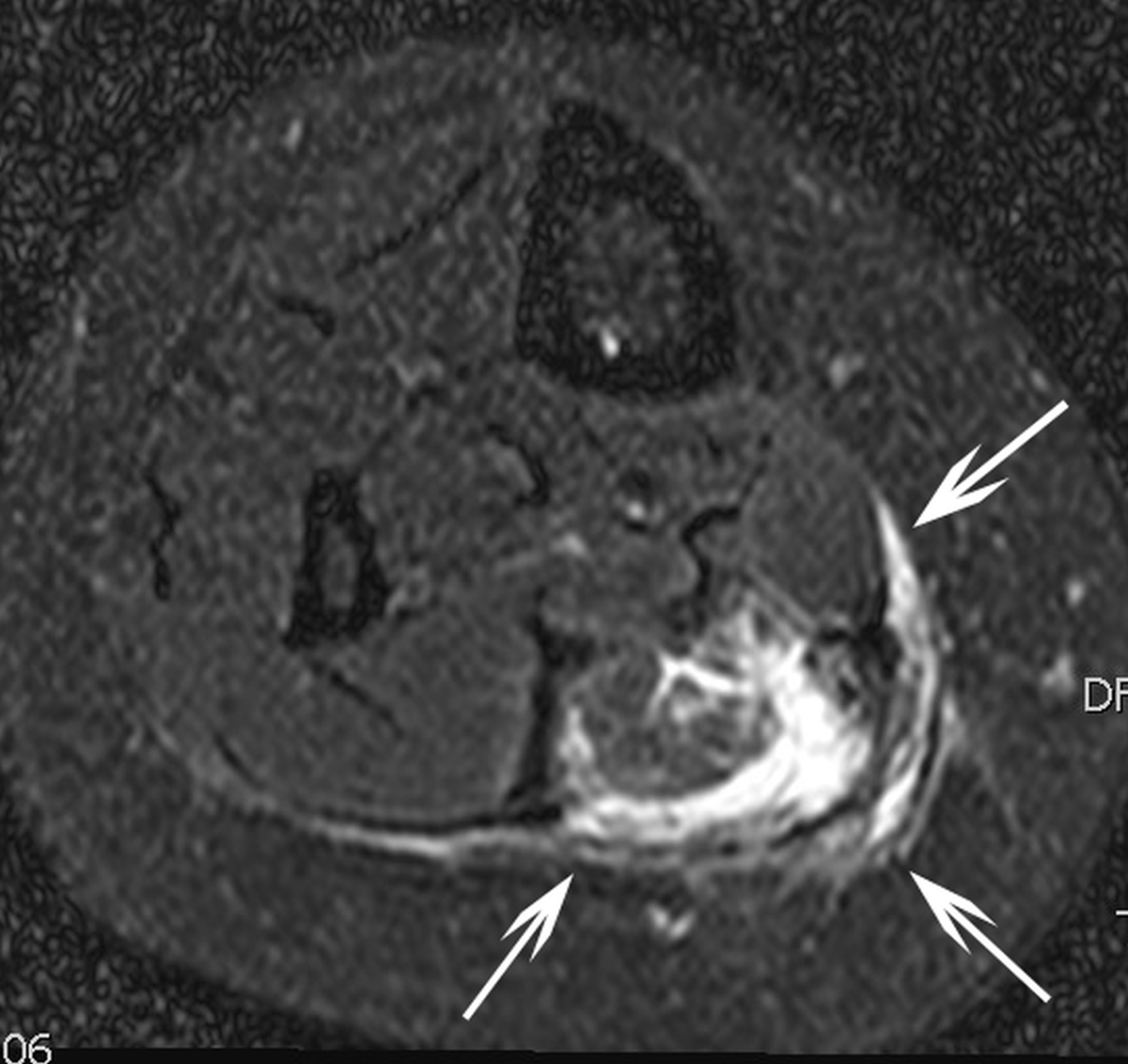
Figure 1b
T2-weigthed axial magnetic resonance image in the same patient three months after percutaneous radiofrequency ablation which was performed after two failed percutaneous sclerotherapies with sotradecol. Hyperintense rim (white arrows) around the ablated area suggestive for perileasional oedema.