
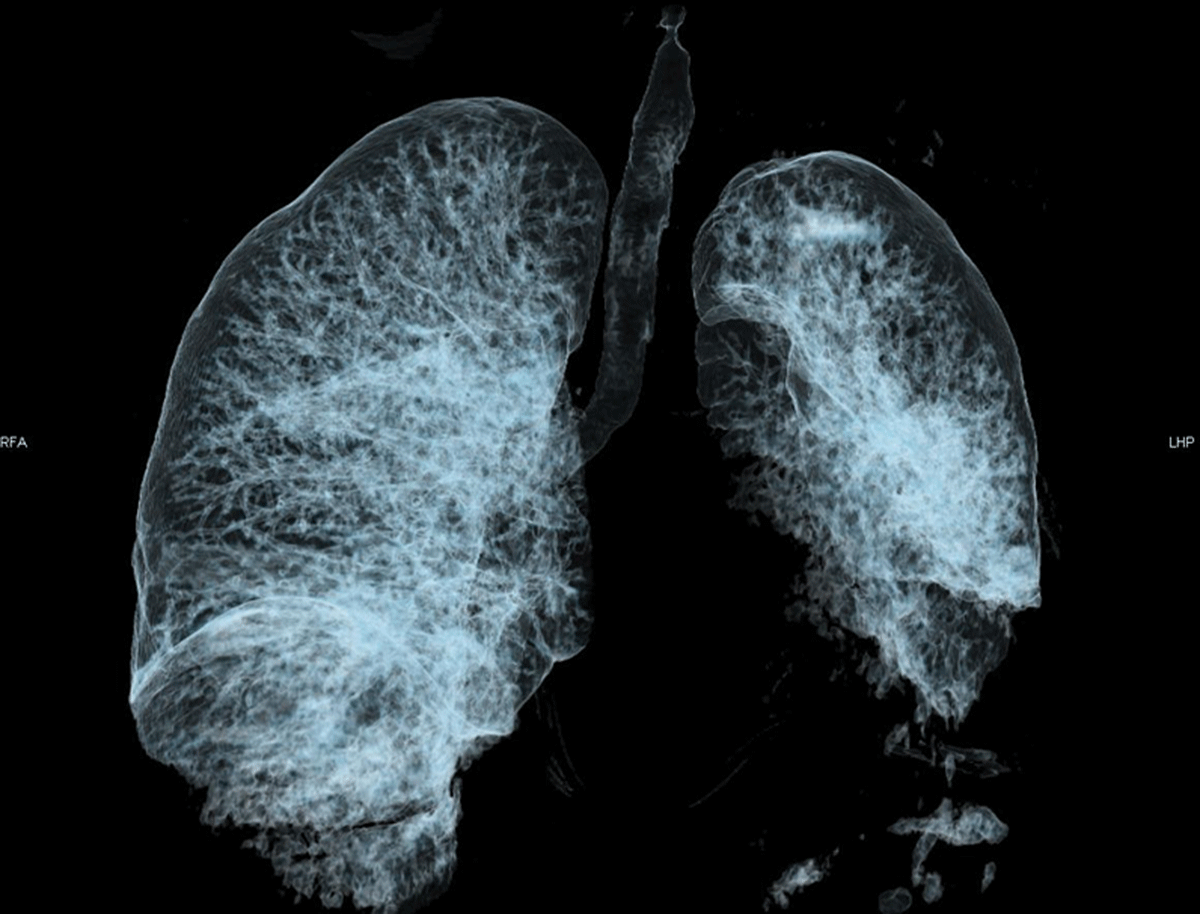
Figure 1
Volume rendering 3D reformat of the airways showing complete obstruction of the non-visible left mainstem bronchus. The air visible in the left lung results from the aerated collapse of the left upper lobe. The left lower lobe suffered non-aerated collapse and thus is not visible.

Figure 2
Axial CT of the pulmonary trunk in mediastinal window. Central bronchi on the left side are outlined by calcifications of bronchial walls cartilage and are filled with mucous (white arrow). The left superior pulmonary vein is not opacified (star). The pulmonary trunk diameter is 38.9 mm (normal being ≤27 mm in females), likely because of PA hypertension.
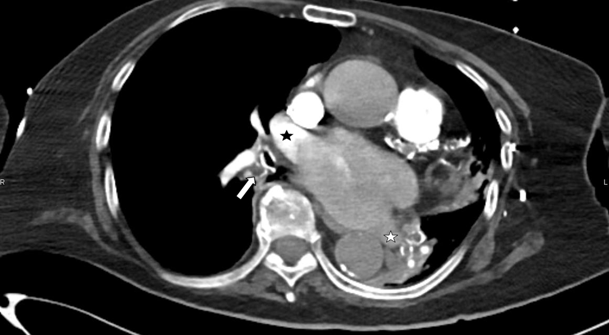
Figure 3
Axial CT of the right lower lobe bronchus that is filled with mucus (arrow), also showing unusual lack of opacification of the left atrium, where only the right superior pulmonary vein is opacified (black star). The left inferior pulmonary vein is not opacified (white star).
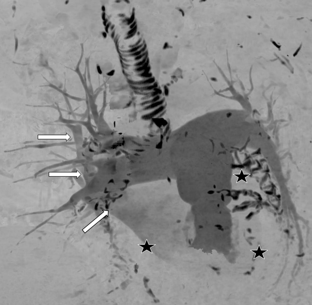
Figure 4
3D maximum intensity projection of all the pulmonary vessels in antero-posterior orientation. The PAs and the right superior pulmonary vein are opacified (arrows). Right lower, left upper, and left lower pulmonary veins are not opacified (stars). The calcified bronchial cartilage and coronary calcifications appear black.