
Table 1
Subject characteristics.
| JUVENILE IDIOPATHIC ARTHRITIS | CONTROL GROUP, PATIENTS WITH FOOT AND ANKLE ARTHRALGIA | p VALUE | |
|---|---|---|---|
| Total number | 165 | 207 | – |
| Gender (% of subgroup) | Male: 64 (38.8%) | Male: 70 (33.8%) | 0.321 |
| Female: 101 (61.2%) | Female: 137 (66.2%) | ||
| Average age (years) | 10.70 | 10.73 | 0.978 |
| [STDEV] | [4.20] | [4.19] | |
| Median (min-max) | 12 (1–18) | 11 (2–18) |
Table 2
Radiographic findings in JIA and non-JIA groups.
| 1. INFLAMMATORY LESIONS | ||||||||
|---|---|---|---|---|---|---|---|---|
| SOFT TISSUE SWELLING | OSTEO-POROSIS | JSN | EROSIONS AND SUBCHONDRAL CYSTS | ANKYLOSIS | MALALIGNMENT | GROWTHABNORMALITIES | PERIOSTEAL BONE FORMATION | |
| JIA (165 patients) | 52 (31.51%) | 24 (14.55%) | 10 (6%) | 15 (9.09%) | 2 (1.21%) | 1 (0.6%) | 1 (0.6%) | 5 (3.03%) |
| non-JIA (207 patients) | 5 (2.41%) | 2 (0.97%) | 0 | 12 (5.79%) | 0 | 0 | 0 | 1 (0.48%) |
| P | p = 0.000 | p = 0.000 | p = 0.000 | p > 0.05 | p > 0.05 | p > 0.05 | p > 0.05 | p > 0.05 |
| 2. NON- INFLAMMATORY LESIONS | ||||||||
| PES PLANUS | HALLUX VALGUS | ANATOMICAL VARIANTS AND BENIGN LESIONS (UNFUSED OSSIFICATION CENTER, TARSAL COALITION, BONE ISLAND, FIBROUS CORTICAL DEFECT, OSTEOCHONDRAL DEFECT) | ASEPTIC NECROSIS | |||||
| JIA (165 patients) | 34 (20.6%) | 11 (6.66%) | 6 (3.63%) | 4 (2.42%) | ||||
| non-JIA (207 patients) | 40 (19.32%) | 20 (9.67%) | 10 (4.83%) | 3 (1.45%) | ||||
| P | p > 0.05 | p > 0.05 | p > 0.05 | p > 0.05 | ||||
[i] * JSN joint space narrowing.
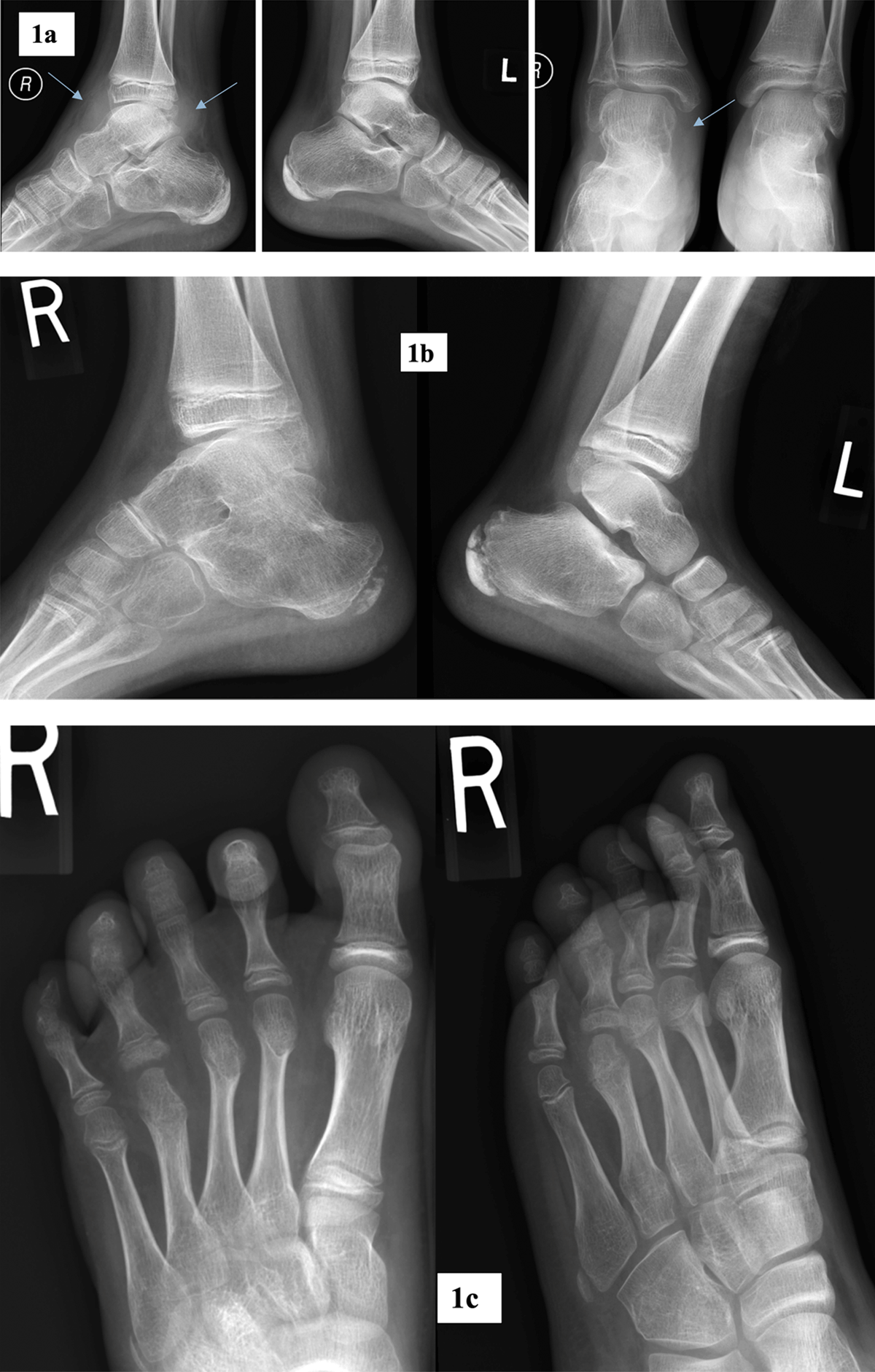
Figure 1
Radiographic abnormalities of foot and ankle.
a. Antero-posterior and lateral radiographs of both ankle joints in a 12-year-old male patient with JIA. Marked soft tissue swelling at the level of right ankle joint (arrows).
b. Lateral radiographs of ankle joints in a 9-years-old male with JIA. Marked osteoporosis of right talar and calcaneal bones with ankylosis in subtalar joint. Talonavicular joint space narrowing with subchondral sclerosis. Normal left ankle joint.
c. Antero-posterior and oblique radiographs of the right foot in a 9-year-old female with JIA. Erosions and subchondral cysts in fourth MTP and PIP joints, periarticular osteoporosis, soft tissue swelling at the level of MTP2 joint.
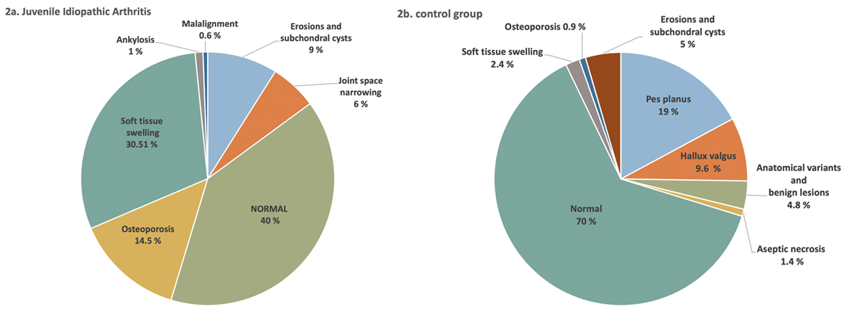
Figure 2
Radiographic findings.
a. In the JIA group. The diagram covers only inflammatory changes. Non-inflammatory lesions, also detected in JIA group, because of overlapping were not included.
b. In the non-JIA group.
Table 3
JIA recognized subtypes.
| JIA SUBTYPES | NUMBER (PERCENTAGE) |
|---|---|
| Oligoarthritis | 76 (46%) |
| Polyarthritis: | 57 (34.54%) |
| RF- negative | 54 |
| RF-positive | 3 |
| ERA (entesitis-related) | 2 (1.21%) |
| Undifferentiated form | 25 (15.15%) |
| Systemic form | 5 (3.03%) |
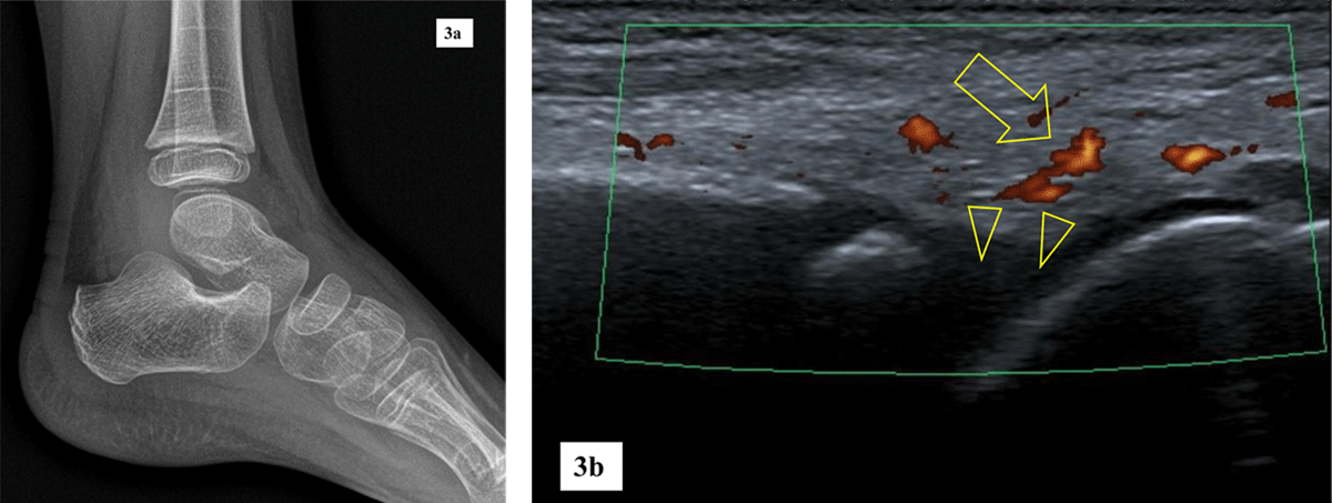
Figure 3
5-year-old girl with confirmed JIA.
a. Lateral radiograph of a left ankle is unremarkable.
b. Sagittal ultrasound of the left ankle shows hypervascular synovium on Power Doppler mode (arrow) and small tibiotalar effusion (arrowheads).
Table 4
Laboratory results.
| JIA NUMBER OF PATIENTS | NON-JIA NUMBER OF PATIENTS | |||
|---|---|---|---|---|
| NEGATIVE | POSITIVE | NEGATIVE | POSITIVE | |
| Antinuclear antibodies (ANA) | 49 | 116 | 72 | 135 |
| Anticyclic citrullinated peptide antibodies (anti-CPP) | 161 | 4 | 207 | 0 |
| Rheumatoid factor (RF) | 162 | 3 | 206 | 1 |
| Human leukocyte antigen (HLA) B-27 antigen | 126 | 39 | 189 | 18 |
| Erythrocyte sedimentation rate level (ESR) | Not elevated in 81 | Elevated in 84 | Not elevated in 161 | Elevated in 46 |
| C-reactive protein (CRP) | Not elevated 120 | Elevated in 45 | Not elevated in 196 | Elevated in 11 |