
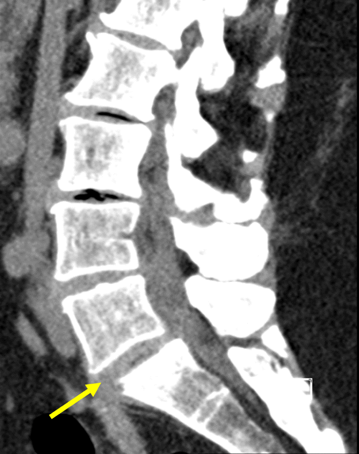
Figure 1
Sagittal non-contrast CT of lumbar spine: Initial CT-scan showing collection from L5-S1 disk with fistulation downwards, an anterolisthesis of L5-S1 with inter-apophysary posterior arthritis and a compression of the right root of S1 by the intervertebral disk.
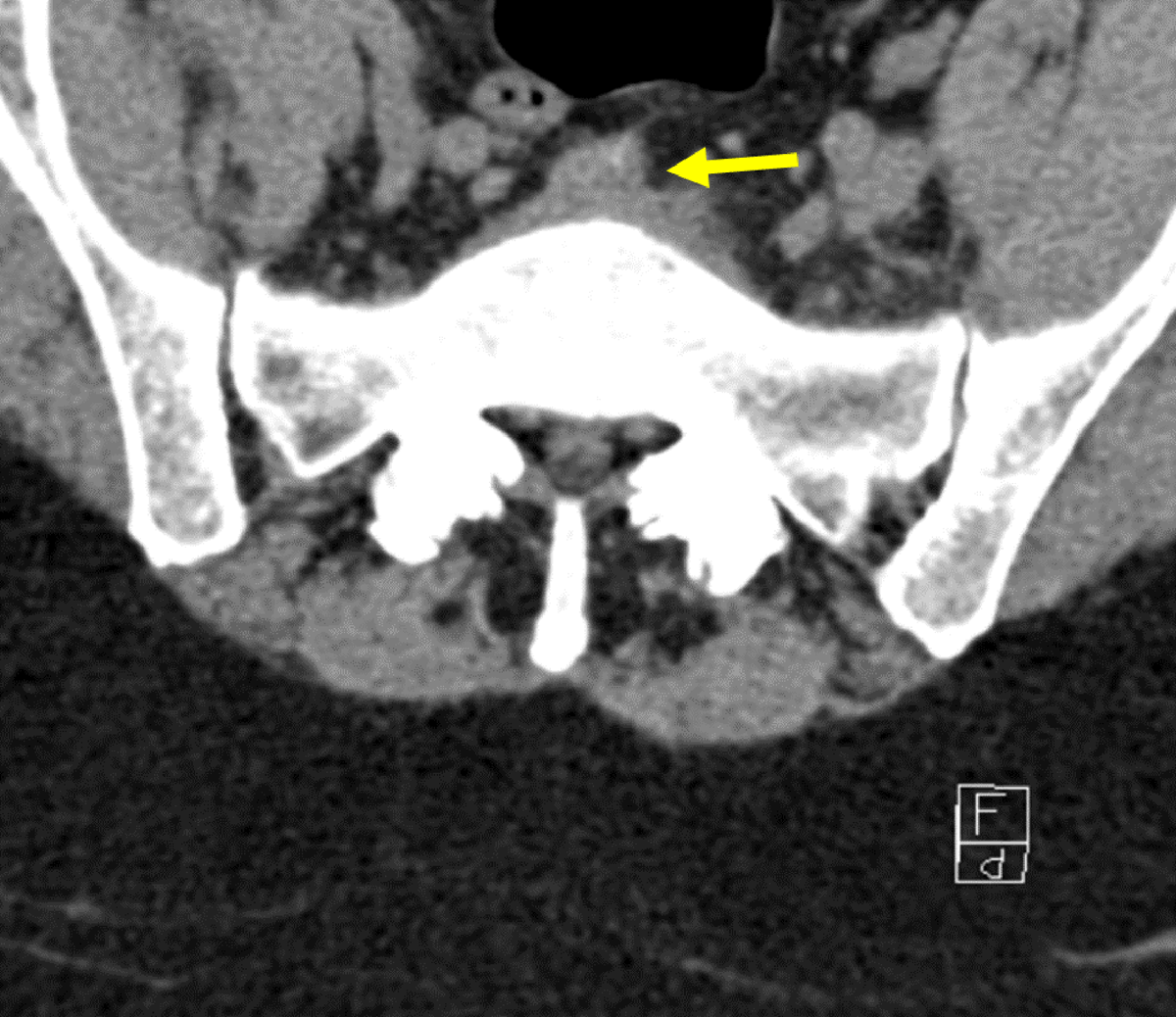
Figure 2
Transverse non-contrast CT of L5-S1 disk: Initial CT-scan showing collection in front of disk.
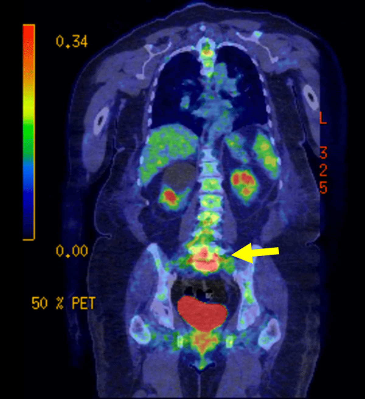
Figure 3
Coronal FDG PET-CT: PET-CT using F-FDG was performed in the context of a possible infection which revealed intense activity at the junction of L5-S1 and the surrounding tissue.
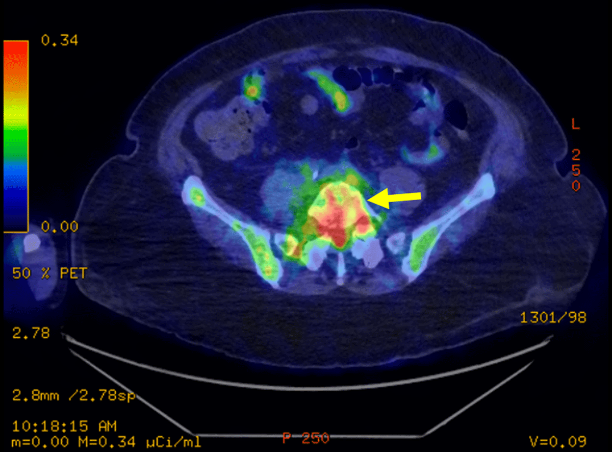
Figure 4
Transverse FDG PET-CT: PET-CT showing hyperfixation of L5-S1 disk.
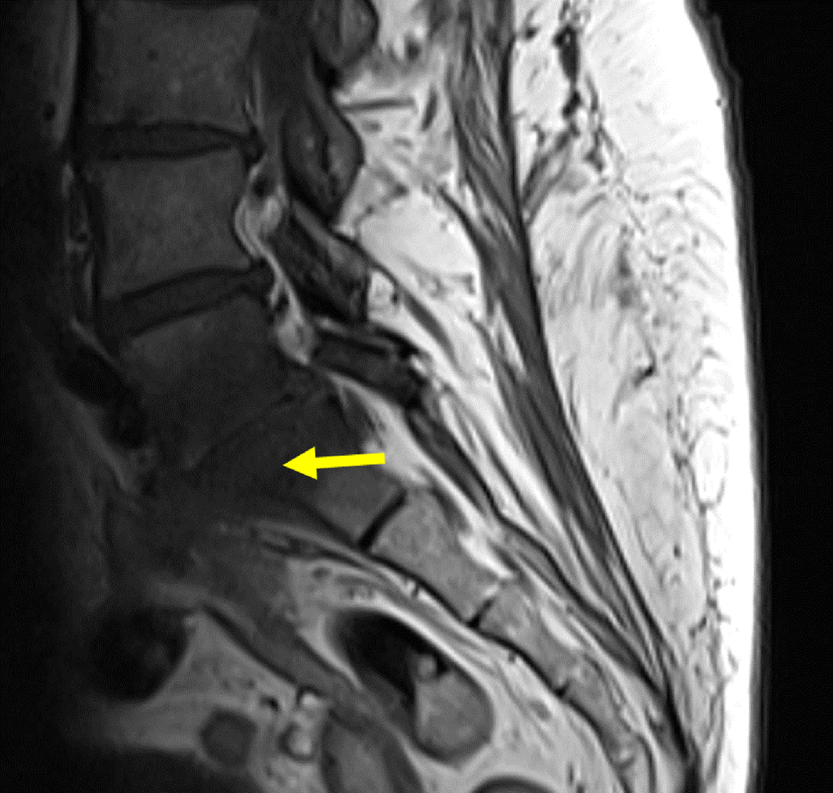
Figure 5
Sagittal contrast MRI in T1 weighting: A medullar bone oedema is seen on either side of the L5-S1 disk. Small collections surround L5-S1 with the largest being 20mm in diameter located on the left psoas muscle with a wall that was intensified by the contrast. This abscess extends posteriorly to the anterior peridural space, the lumbar vertebral bodies, and the last thoracic vertebra.
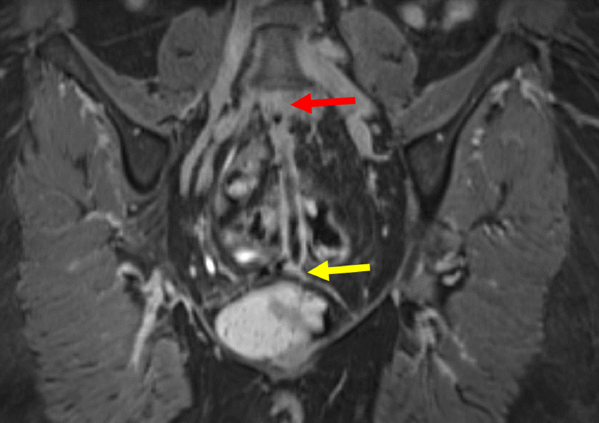
Figure 6
Coronal 3D contrast MRI in T1 fat saturation weighing: Full path of fistula from L5-S1 (yellow arrow) to the vagina (red arrow). The fistula presents with a hyperintense wall surrounding a hypointense lumen.
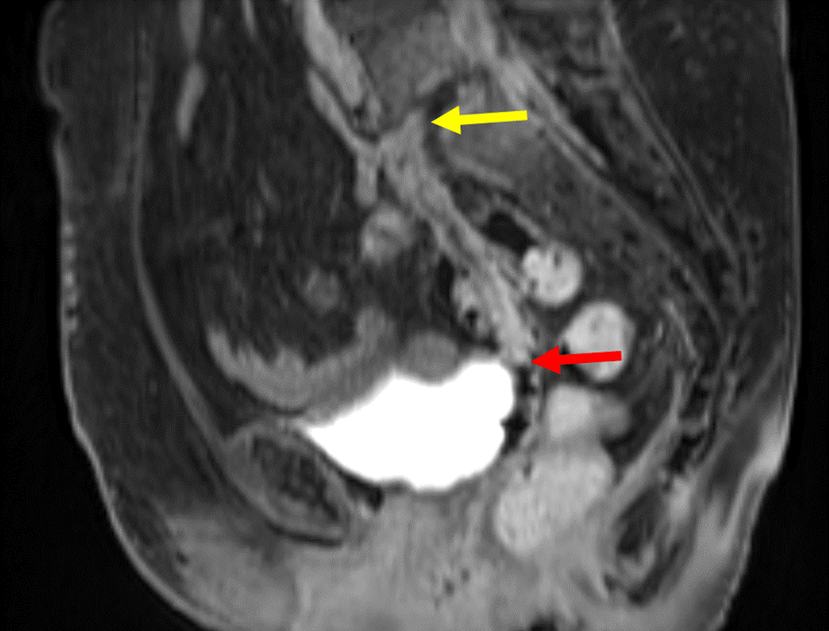
Figure 7
Sagittal contrast MRI in T1 fat saturation weighing: Full path of fistula from L5-S1 (yellow arrow) to the vagina. The fistula (red arrow) presents with a hyperintense wall surrounding a hypointense lumen.
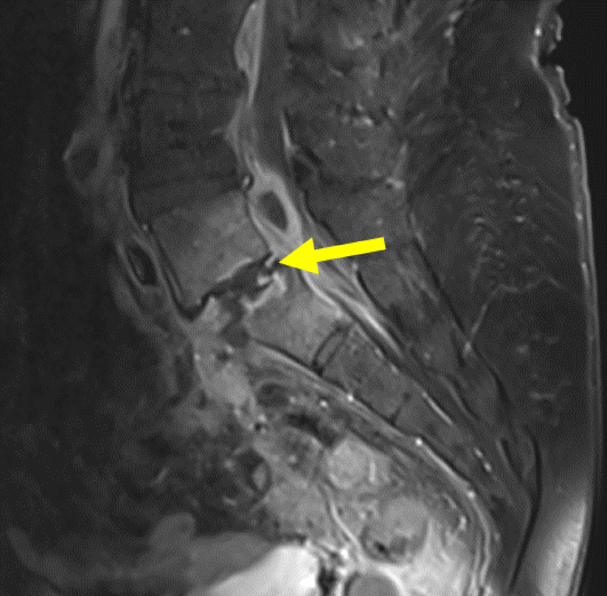
Figure 8
Sagittal contrast MRI in T1 fat saturation weighing: Hyperintense L5-S1 vertebrae and surrounding tissue, showing spondylodiscitis (yellow arrow). There is slight anterolisthesis of L5 upon S1.
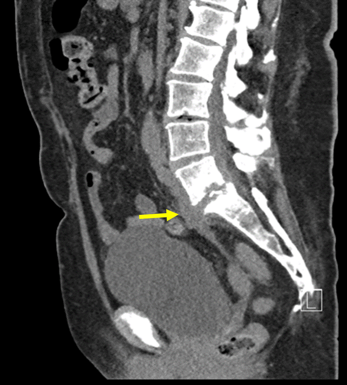
Figure 9
Sagittal contrast CT: Contrast CT-scan showing spondylodiscitis of L5-S1, with infiltration into the soft tissue surrounding S1. Additionally, a 9.7cm fistula can be seen, starting at the intervertebral disk of L5-S1 (yellow arrow) to the vagina.
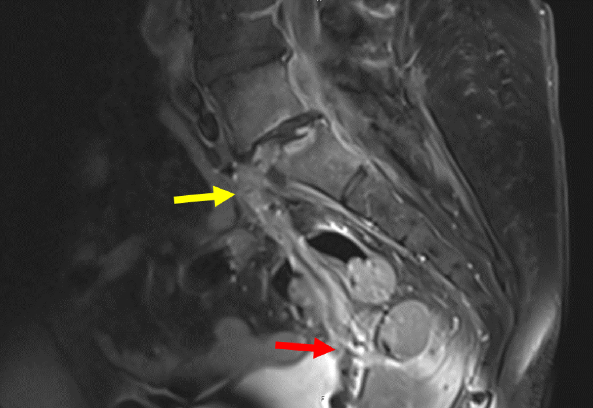
Figure 10
Sagittal contrast MRI in T1 weighting: Pre-operative MRI showing an epidural collection behind the vertebral body of L5 and within the L5-S1 disk (yellow arrow), fistulising into the vagina (red arrow).