
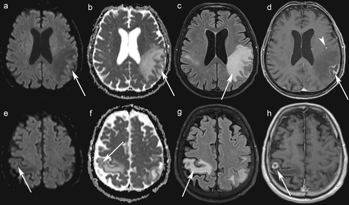
Figure 1
Brain MRI. Axial Diffusion-Weighted Imaging (b = 1500 s/mm²) show no increased signal in the left and right parietal lobe lesions (a, e) while axial ADC maps show a greater signal than that of the unaffected white matter (b, f) (arrows). Axial FLAIR images show edema around the lesions (c, g). Axial contrast-enhanced T1-WI shows rim-enhancing lesions in the left and the right parietal lobes (arrows) (d, h), and one punctate lesion in the left frontal lobe (arrowhead) (h).
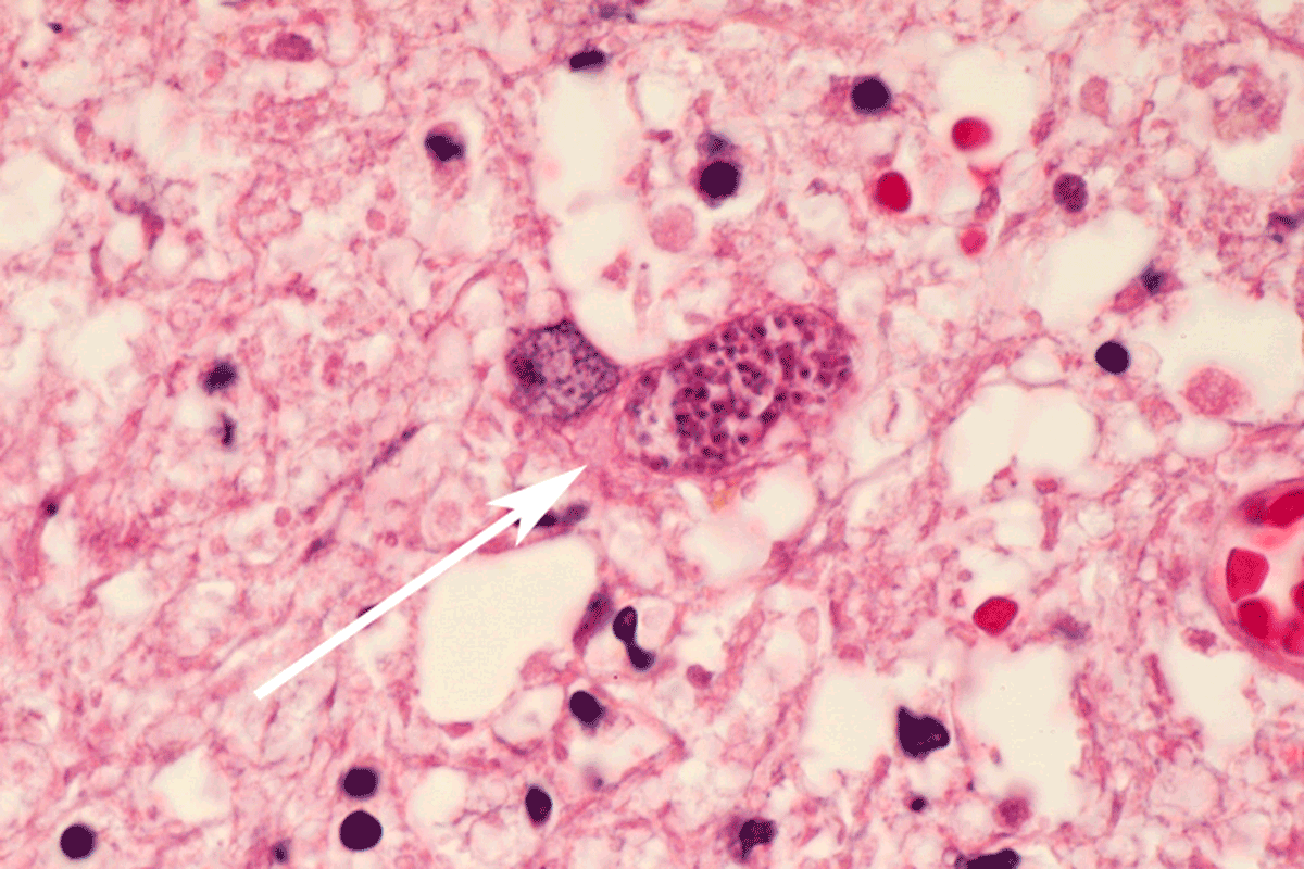
Figure 2
Histological examination (hematoxylin and eosin stain, ×100) shows Toxoplasma gondii pseudocyst consisting of multiple protozoa within a cell (arrow).