
Table 1
Indications for main splenic artery embolization.
| INDICATION | NUMBER | % |
|---|---|---|
| Splenic rupture | 22 | 34% |
| Traumatic | 12 | 18% |
| Coagulopathy | 4 | 6% |
| Leukaemia | 2 | 3% |
| Postoperative | 2 | 3% |
| Pancreatitis | 1 | 1.5% |
| Idiopathic | 1 | 1.5% |
| Pseudoaneurysm | 19 | 29% |
| Pancreatitis | 15 | 23% |
| Postoperative | 3 | 5% |
| Traumatic | 1 | 1.5% |
| Aneurysm | 12 | 18% |
| Focal extravasation from main splenic artery | 9 | 14% |
| Postoperative hemorrhage | 6 | 9.5% |
| Pancreatitis | 3 | 5% |
| Splenomegaly | 2 | 3% |
| Preoperative | 1 | 1.5% |
Table 2
Anatomic characteristics and associated symptoms of the (pseudo)aneurysms.
| ANEURYSM | PSEUDOANEURYSM | |
|---|---|---|
| Total number (n = 31) | 12 (38.7%) | 19 (61.3%) |
| Symptomatic patient | 2 (17%) | 6 (32%) |
| Location | ||
| Proximal MSA | 2 (17%) | 2 (11%) |
| Middle MSA | 5 (42%) | 9 (47%) |
| Distal MSA | 5 (42%) | 8 (42%) |
| Mean diameter (mm) | 30.5 mm (16 mm–45 mm) | 41.1 mm (22.7 mm–59.5 mm) |
[i] MSA: main splenic artery.
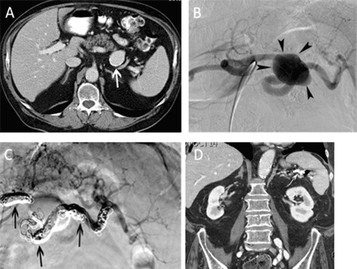
Figure 1
Main splenic artery embolization for atherosclerotic aneurysm. (A) Contrast-enhanced CT-scan in a 74-year-old man revealed an asymptomatic, atherosclerotic aneurysm (white arrow) with a maximal diameter of 37 mm. The total splenic volume was 418 ml. (B) Selective angiography of the celiac trunk confirmed the saccular aneurysm (arrowheads) in the middle third of the main splenic artery. (C) Completion angiography after coil embolization (arrows) demonstrated exclusion of the aneurysm and reinjection of the intrasplenic arteries through gastric collaterals. (D) Follow-up CT-scan 5 years after coil embolization revealed an homogeneously enhancing spleen with a total volume of 264 ml.
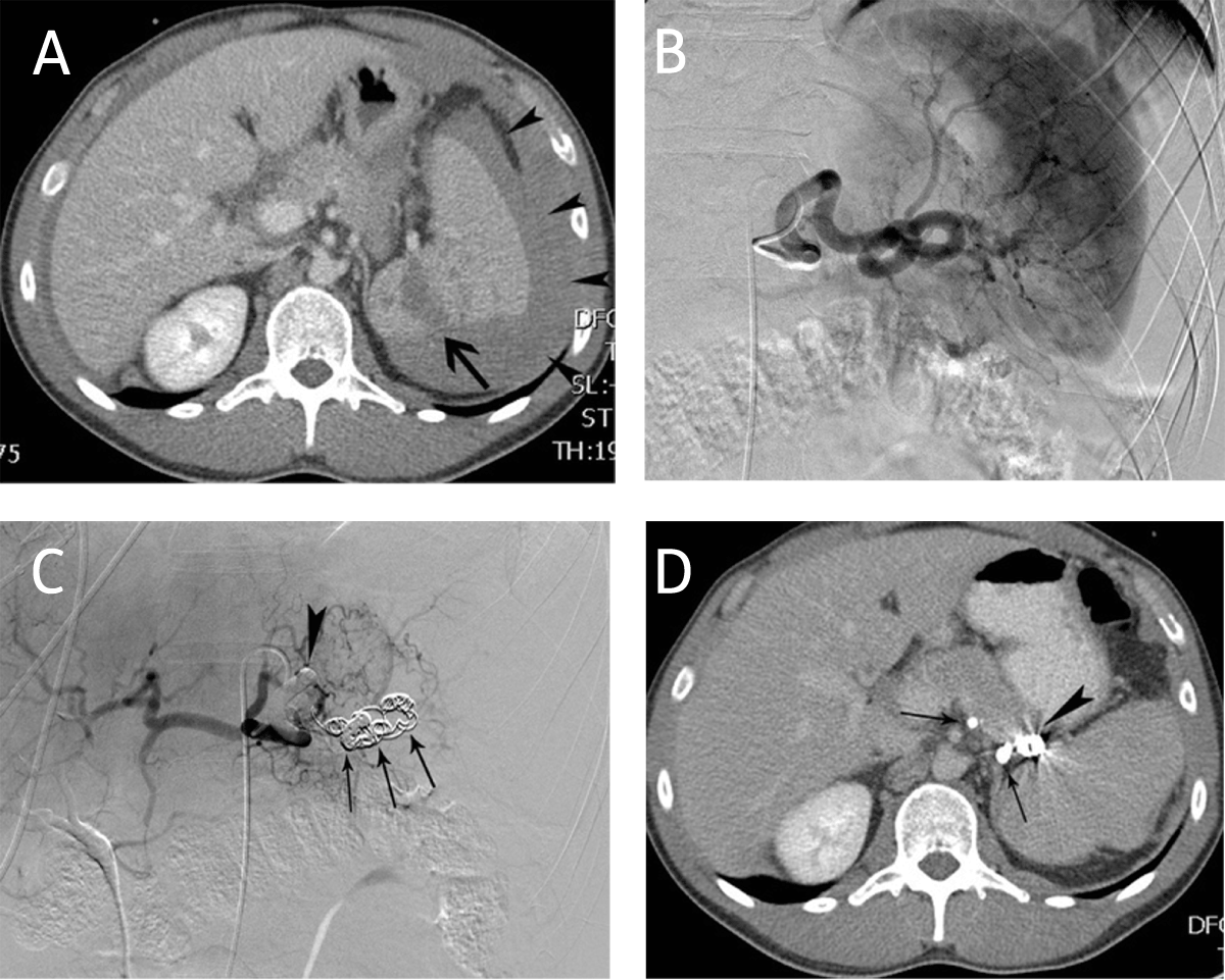
Figure 2
Main splenic artery embolization for trauma. (A) CT-scan in a 27-year-old man after a traffic accident revealed splenic laceration (arrow) and a perisplenic hematoma (arrowheads). The volume of the spleen was 270 ml. (B) Corresponding selective splenic angiography did not reveal contrast extravasation. (C) Main splenic artery embolization was performed with coils (arrows) and glue (arrowhead). (D) Follow-up CT-scan 8 months later demonstrated the glue (arrows) and coils (arrowhead) in the splenic artery. The total splenic volume was 290 ml.
Table 3
Distribution of embolics among indication for embolization.
| ETIOLOGY OF SPLENIC ARTERY DISEASE | ||||||
|---|---|---|---|---|---|---|
| ANEURYSM | PSEUDO- ANEURYSM | SPLENIC RUPTURE | HEMORRHAGE | SPLENOMEGALY | OTHER | |
| Embolic material | ||||||
| Glue | 1 | 3 | 2 | 1 | 0 | 0 |
| Glue + microcoils | 0 | 2 | 1 | 2 | 0 | 0 |
| Microcoils | 9 | 13 | 19 | 6 | 1 | 1 |
| Microcoils + microparticles | 2 | 1 | 0 | 0 | 1 | 0 |
Table 4
Follow-up radiological modality after main splenic artery embolization.
| BEFORE EMBOLIZATION | AFTER EMBOLIZATION | |||
|---|---|---|---|---|
| N | % | N | % | |
| Computed tomography (CT) | 59 | 90.7% | 26 | 40% |
| US | 4 | 6.1% | 3 | 4% |
| Magnetic resonance imaging (MRI) | 2 | 3.1% | 2 | 3% |
Table 5
Imaging follow-up of enhancing splenic volume in patients with a pre-interventional normal spleen (group 1) versus patients with a traumatic splenic rupture (group 2).
| TRAUMATIC SPLENIC RUPTURE (n = 16) | SPLENIC ARTERY (PSEUDO)ANEURYSM (n = 7) | |
|---|---|---|
| Pre-interventional splenic volume | 261 ml | 311 ml |
| Post-interventional splenic volume | 215 ml | 257 ml |
| Median follow-up (days) | 702 (43–2095) | 1163 (61–3946) |
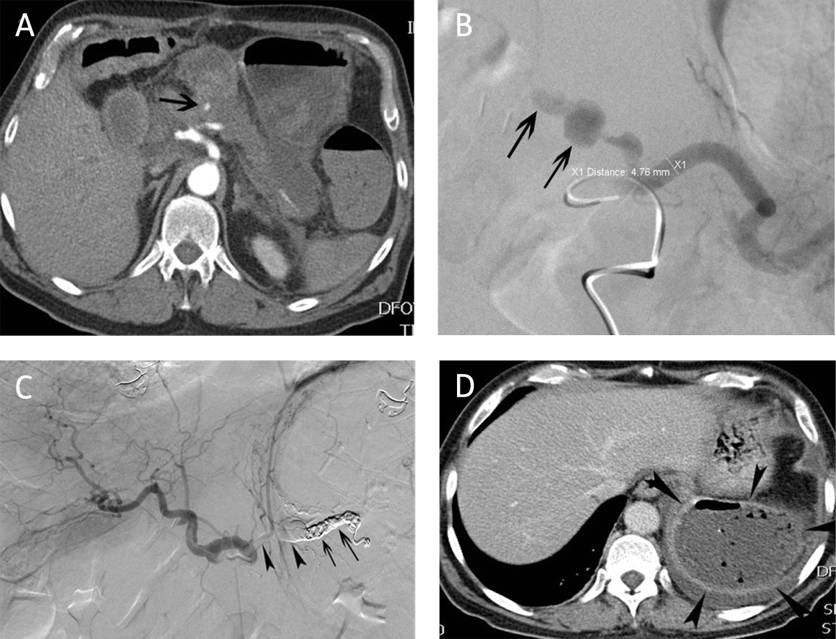
Figure 3
Main splenic artery embolization for postoperative hemorrhage. (A) CT-angiography in a 64-year-old man 19 days after pancreatico-duodenectomy revealed contrast extravasation (arrow) in the proximal part of the main splenic artery. (B) Corresponding selective splenic arteriography confirmed the contrast extravasation and a pseudoaneurysm (arrows). (C) Embolization of the hemorrhage was performed with a combination of coils (arrows) and glue (arrowheads). (D) CT eight days after embolization revealed multiple air bubbles in a completely necrotic splenic tissue compatible with abscess.