

Figure 1
A 56-year-old man presented with status epilepticus and coma four days after bi-pulmonary graft. (A) Non-enhanced CT shows ventricular and sulcal effacement suggesting brain edema. (B) Fluid-attenuated inversion recovery (FLAIR) sequence shows bilateral cortical swelling and high signal, with sparing of the occipital cortex, as well as signal abnormalities in both caudate nuclei and thalami. (C) Diffusion weighted-imaging (DWI; b-value = 1000 s/mm2) and (D) apparent diffusion coefficient (ADC) map show respectively high and low signal of the cortex at the same levels, consistent with cytotoxic edema. (E) DWI sequence at a higher level shows sparing of the peri-rolandic cortex. (F) Follow-up FLAIR imaging at four weeks shows development of atrophy and gliosis in insula bilaterally.
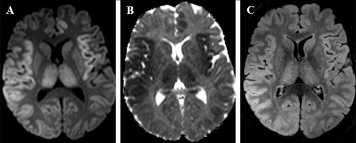
Figure 2
A 45-year-old man presented with seizures and fulminant hepatitis with hepatic failure, secondary to acetaminophen intoxication. (A) DWI (b-value = 1000 s/mm2) and (B) ADC map show cortical signal abnormalities in bilateral insulas and parietal lobes, left cingulate gyrus and both thalami, respectively high and low signal, consistent with cytotoxic edema. (C) FLAIR sequence shows high signal in the right parietal cortex and bilateral thalami.
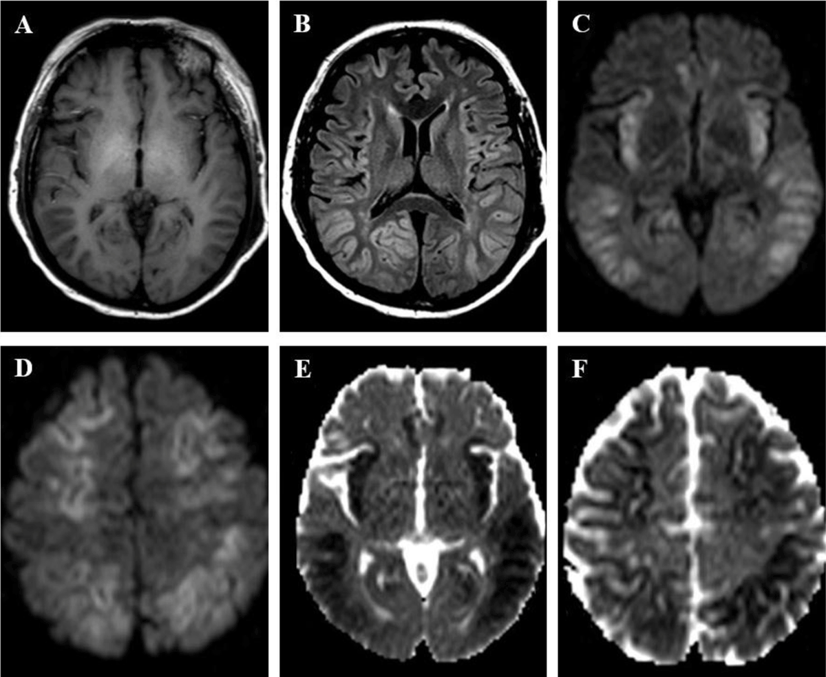
Figure 3
A 49-year old man was found in a coma. Clinical and laboratory findings indicated decompensated cirrhosis with encephalopathy. (A) T1-weighted imaging shows high signal in globi pallidi, suggesting underlying chronic hepatic disease. (B) FLAIR image shows bilateral and symmetric signal abnormalities in insular, cingulate, frontal and parietal cortices. (C, D) DWI (b-value = 1000 s/mm2) and (E, F) ADC show respectively high and low signal in affected cortices, consistent with cytotoxic edema, with sparing of occipital and peri-rolandic cortices.