
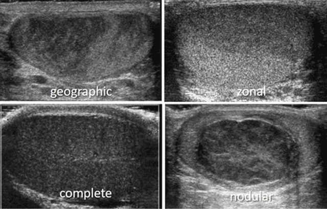
Figure 1
Geographic, zonal, complete or nodular decrease of testicular echogenicity.
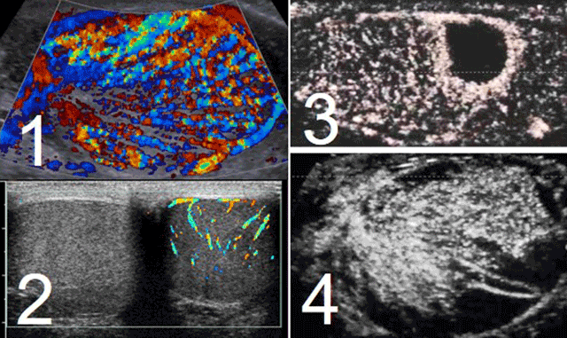
Figure 2
Testicular vascular analysis: hypervascularization at CD in case of acute orchitis (1), absence of CD signal in case of acute torsion (2), typical absence of perfusion at CEUS in case of segmental ischemia (3) or in case of trauma (4).
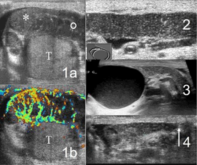
Figure 3
Excretory tube analysis: typical acute epididymitis (1a, 1b), dilatation of the epididymal body (2), cyst of the epididymal head (3), absence of vas deferens (4).