
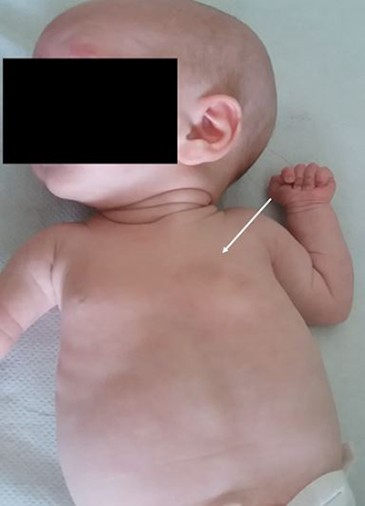
Figure 1
Photograph of the baby girl at 2 months of age demonstrating the asymmetry in the chest wall with impression of missing muscle and ribs 4 and 5 on the left side (white arrow). The bilateral gynaecomastia is also visible.
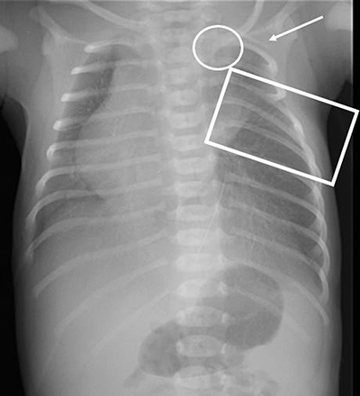
Figure 2
Chest X-ray at day 0 shows 11 ribs on the right side and 11 ribs plus an apparent cervical rib on the left side (white arrow). The intercostal spaces between ribs 4 and 6 on the left side appear narrowed (rectangle). The medial end of the left clavicle (circle) has an abnormal low position compared to the right clavicle and the cardiac silhouette is positioned on the right side.
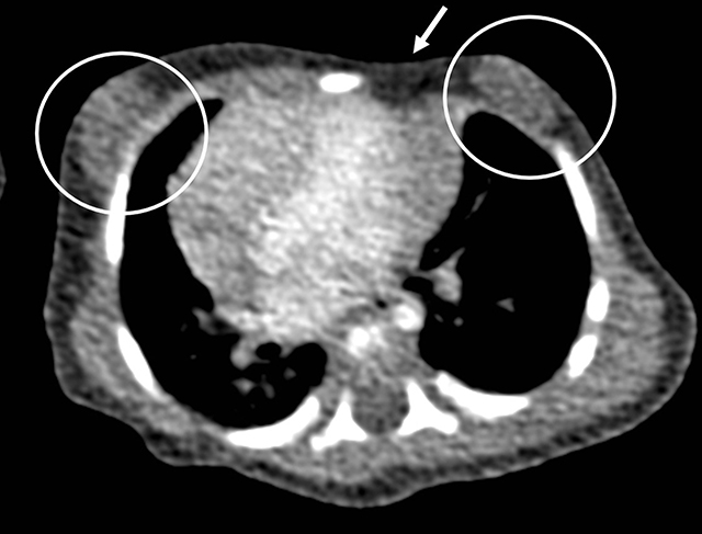
Figure 3
Axial CT-scan image in mediastinal window shows absence of the sternocostal head of the left major pectoral muscle (white arrow) with subsequent depression in the left chest wall. There is bilateral gynaecomastia (circles). The heart is positioned more to the right side than normal.
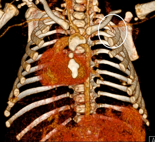
Figure 4
3D reconstruction of the chest CT-scan shows a dysplastic left sternal half and a dextroposition of the heart. There is a hypoplastic first rib on the left side (circle).
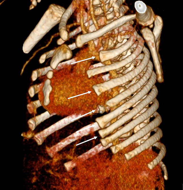
Figure 5
3D reconstruction of the chest CT-scan shows partial agenesis of the anterior arches of ribs 2, 4 to 7 on the left side (white arrows).
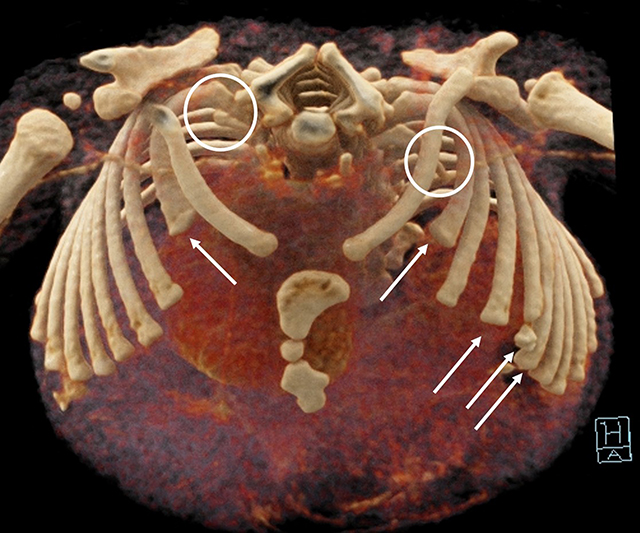
Figure 6
3D reconstruction of the chest CT-scan shows bilateral hypoplastic first ribs (circles) and a partial agenesis of the anterior arches of ribs 2 bilaterally and of ribs 4 to 7 on the left side (white arrows).