
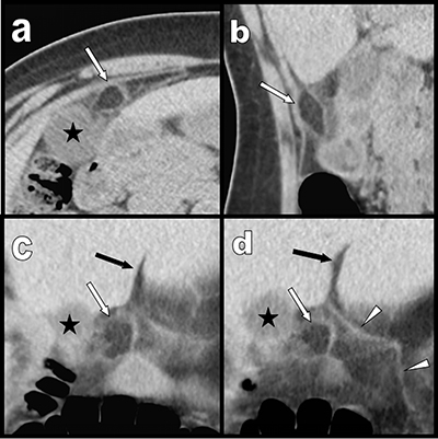
Figure 1
Case 1: transverse (a), sagital (b) and coronal oblique (c) and (d) multiplanar reconstructions show a pre-hepatic area of fat stranding surrounding a 1,5 cm well demarcated inflammatory lipomatous process (white arrows) just under the hepatic fissure (black arrow) and in very close vicinity of the right edge of the ligamentum teres (white arrowheads). Black star = gallbladder.
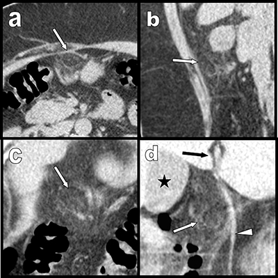
Figure 2
Case 2: similarly to case 1, transverse (a), sagital (b) and coronal oblique (c) and (d) multiplanar reconstructions also show a pre-hepatic area of fat stranding surrounding a 1,5 cm inflammatory lipomatous mass (white arrows) just under the hepatic fissure (black arrow) and in very close vicinity of the ligamentum teres (white arrowheads). Black star = gallbladder. The background noise is more marked than in case one and is related to the obesity of the second patient.