

Figure 1
Axial contrast-enhanced CT: A biconcave lesion lining the superolateral left orbital wall.
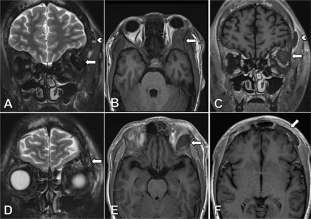
Figure 2
Coronal TSE T2-weighted image with fat suppression (A) shows a markedly hypointense lesion located in the lateral aspect of the left orbital roof (arrow) and a second lesion lining the external table of the frontal bone on the left side (arrow head). On axial TSE T1-weighted images (B) the lesion exhibits a isointense signal compared to the adjacent bone, without lesional enhancement after injection of gadolinium (E). Coronal contrast-enhanced TSE T1-weighted image (C) reveals a faint perilesional enhancement, more prominently in the lesion lining the frontal bone on the left (arrow head). Coronal TSE T2-weighted image with fat suppression (D) shows asymmetrical, discrete bone edema and prominent nonenhancing lesions on contrast-enhanced TSE T1-weighted image (F) in the frontal bone on the left side compared to the right side.