
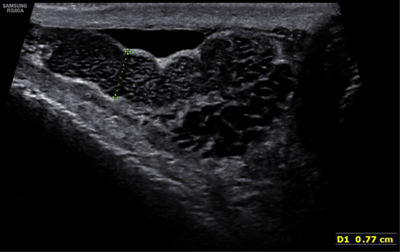
Figure 1
Enlargement of the right epididymis with multiple small cystic dilatations.
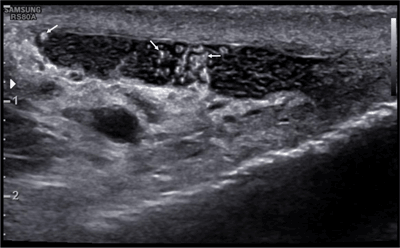
Figure 2
Irregularly shaped, hyperechoic particles (arrows) within the cystic dilatations of the epididymis.
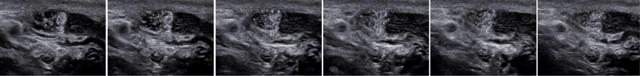
Figure 3
Selected images of an ultrasound film. Oscillating movement of the hyperehoic particles.