
Figure 1
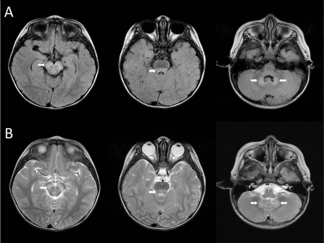
FLAIR (A) and T2WI (B) demonstrate hyperintense lesions in the midbrain, dorsal pons, and cerebellar dentate nuclei (white arrows).
Table 1
Clinical characteristics, virological data, and MRI findings of the five patients with coxsackievirus encephalitis in the current case and the literature.
| Patient no. | Sex | GA (weeks) | Age at onset of illness (days/PMA in weeks) | MRI Findings | Serotype | Reference |
|---|---|---|---|---|---|---|
| 1 | M | 35 | 3/35 | Periventricular white matter | CVB1 | [3] |
| 2 | F | 36 | 3/36 | Periventricular white matter | CVB1 | [3] |
| 3 | F | Term | 20/NA | Dorsal pontomedullary junction, right basal ganglion | CVB1 | [1] |
| 4 | F | 37 | 12/39 | Internal capsule, corpus callosum, cerebral deep and subcortical white matter | CVB2 | [2] |
| 5 | F | NA | 2 years old | Midbrain, dorsal pons, and cerebellar dentate nuclei | CVB3 | Current case |
[i] GA: gestational age; NA: not available; PMA: postmenstrual age.