
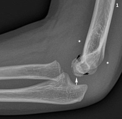
Figure 1
Conventional radiography of the right elbow, lateral view. The capitellum has a slightly irregular articular contour (white arrow) and there is a radiolucent line in the subchondral bone (black arrows). Note slight joint effusion with displacement of the elbow fad pads (asterisks).
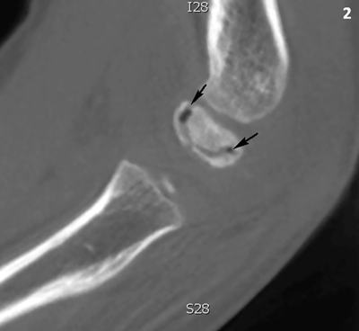
Figure 2
Cone beam CT of the right elbow, sagittal reformatted image. The capitellum has an increased density. There is a subchondral crescent-shaped vacuum phenomenon (black arrows).