Have a personal or library account? Click to login
Temporal Bone CT Cover

Figures & Tables

jbsr-102-S1-1643-g1.png
Figure 1

On this axial CT image of a temporal bone at the level of the oval window opacification of the tympanic cavity is seen, with complete erosion of the stapes and incus long process. The malleus neck is intact (white arrow): cholesteatoma with ossicular chain erosion.
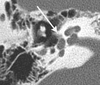
jbsr-102-S1-1643-g2.png
Figure 2

On this axial CT image of a temporal bone at the level of the oval window, the otic capsule appears hypodense in the region of the fissula antefenestram (white arrow): fenestral otosclerosis.

DOI: https://doi.org/10.5334/jbsr.1643 | Journal eISSN: 2514-8281
Language: English
Submitted on: Sep 4, 2018
Accepted on: Sep 26, 2018
Published on: Nov 17, 2018
Published by: Ubiquity Press
In partnership with: Paradigm Publishing Services
Publication frequency: 1 issue per year
Keywords:

© 2018 Marc Lemmerling, published by Ubiquity Press
This work is licensed under the Creative Commons Attribution 4.0 License.