
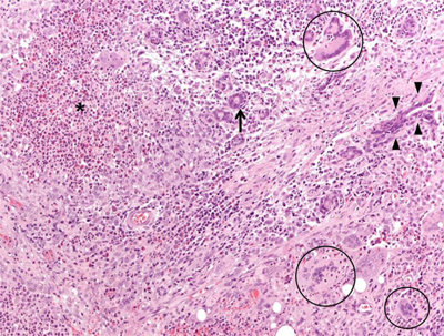
Figure 1
Microphotograph (magnification: 40X) showing a granulomatous mastitis. Mammary parenchyma is characterized by the presence of dense necrotic foci (asterisk), giant multinucleated cells (circles), and normal breast ducts in transversal (arrow) and longitudinal section (arrowheads), trapped by neutrophilic-eosinophilic infiltrate.
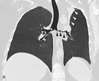
Figure 2
High-resolution computed tomography of the chest, coronal reconstruction, showing a marked wall thickening of the right main bronchus (asterisk), stenosis of the left main bronchus (black arrowheads), and segmental atelectasis in the upper lobe of the left lung (white arrowheads).
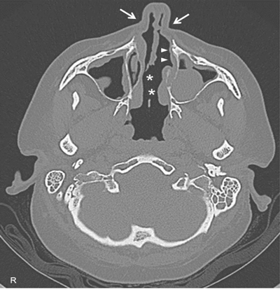
Figure 3
High-resolution computed tomography of paranasal sinuses, axial projection, showing a perforation of the nasal septum (asterisks), the absence of the anterior half of the left inferior turbinate (white arrows), and the atrophy of the upper lateral cartilages (white arrowheads).