
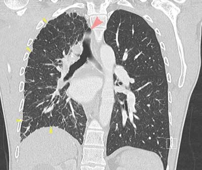
Figure 1
CECT of the thorax, lung window, coronal reformatted image. Hyperinflation of the left lung, smaller volume of the right lung. Mediastinal shift to the right side (red arrow pointing to displaced trachea). Thickened intralobular and interlobular septa (yellow arrows).
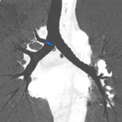
Figure 2
CECT of the thorax, lung window, coronal MinIP reconstruction. Tracheal bronchus arising at the right side of the carina (blue arrow).
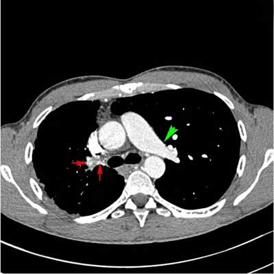
Figure 3
CECT of the thorax, soft tissue window, axial image. Normal appearance of the left pulmonary artery (green arrow). Absent right pulmonary artery. Prominent bronchial arteries supplying the right lung (red arrows). Smaller volume of the right lung.