
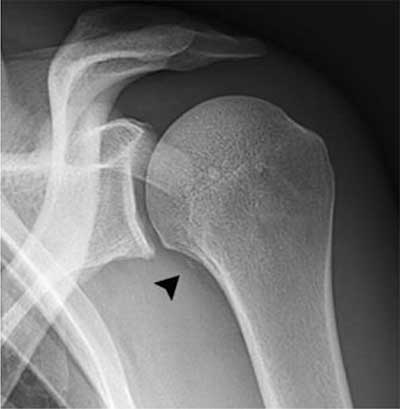
Figure 1
A 29-year-old rugby player presenting after shoulder trauma. Standard radiography of the left shoulder. A small fleck of bone (black arrowhead) is seen medial to the proximal humerus.
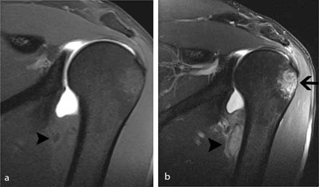
Figure 2
MR arthrography of the left shoulder performed four weeks after initial injury. a) On the coronal FS T1-WI, there is an extra-articular lesion with slightly inhomogeneous signal intensity (black arrowhead) at the medial side of the humeral diaphysis. b) The lesion (black arrowhead) is better seen on the FS T2-WI. Note also the posttraumatic bone marrow edema (black arrow) at the superolateral aspect of the humeral head.
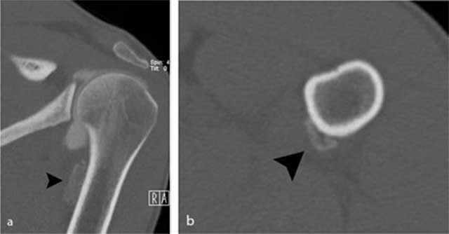
Figure 3
Additional CT images of the proximal humerus. a) Oblique coronal reformatted images and b) axial reformatted images reveal the presence of an extra-articular calcification (black arrowhead) near the insertion of the teres major tendon.
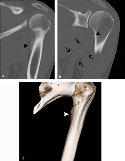
Figure 4
Follow-up CT five weeks later. a) Oblique coronal reformatted image of the proximal humerus shows a slightly more dense aspect of the periphery of the calcification (black arrowhead). b) MIP reconstructions (1.5 mm) clearly show that the calcification is located at the medial crest of the lesser tuberosity (black arrowhead). Note also the perimuscular fat pads surrounding the teres major muscle (black arrows). c) Volume Rendering Technique (VRT) image revealing irregular contour (white arrowhead) near the medial crest of the lesser tuberosity.
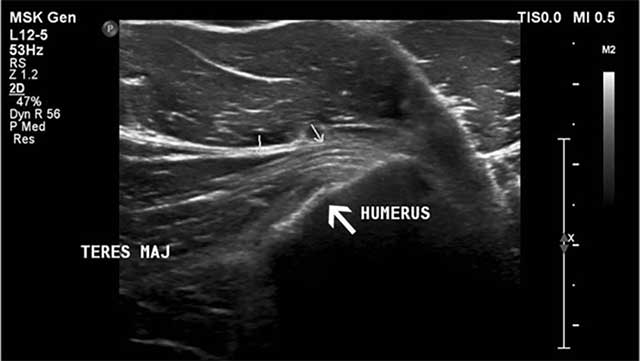
Figure 5
Ultrasound imaging (axial image) reveals an irregular delineation (large white arrow) of the teres major insertion at the medial aspect of the humerus posteromedial to the pectoralis major tendon (small arrows).
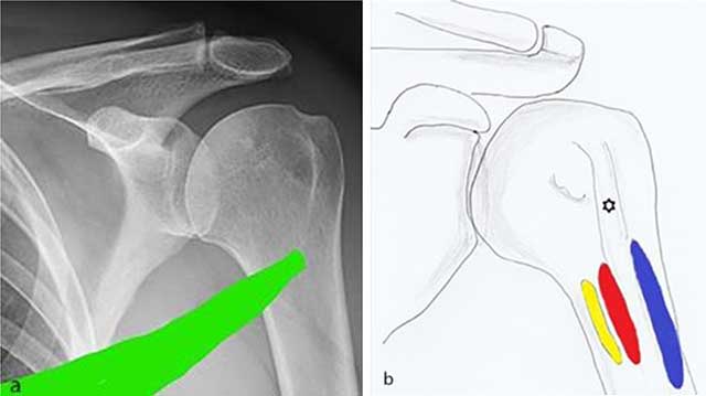
Figure 6
a) Spiral anatomical course of the teres major which may explain shearing forces leading to the periosteal stripping is demonstrated. b) Anatomical drawing showing the close anatomical relationship of the insertion of the teres major (yellow), latissimus dorsi (red) and pectoralis major (blue) tendon.