
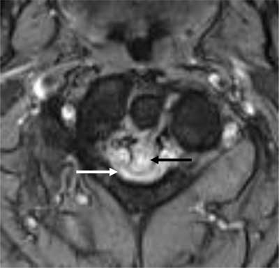
Figure 1
Axial T2-weighted image. The retro-odontoid pseudotumor is seen as a hyperintense mass (black arrow) which extrudes through the transverse ligament and compresses the myelum (white arrow).
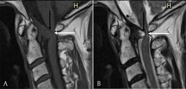
Figure 2
(A) Sagittal T1-weighted image. The retro-odontoid pseudotumor is seen as a mass (black arrow) extruding through the transverse ligament and compressing the myelum (white arrow). It has an isointense signal compared to the myelum. (B) Sagittal T2-weighted image. The retro-odontoid pseudotumor has a hyperintense signal (black arrow) and compresses the myelum (white arrow).
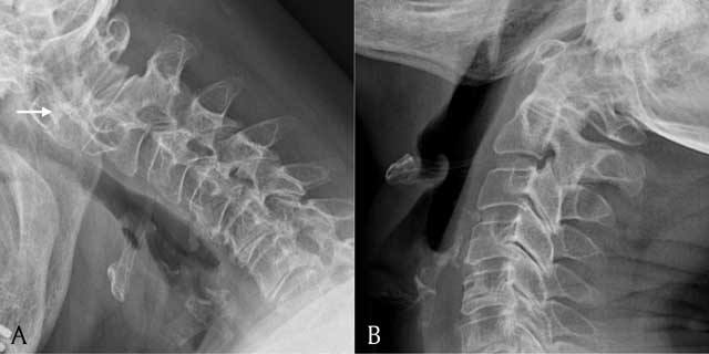
Figure 3
A = lateral view of the cervical spine in flexion; B = lateral view of the cervical spine in extension. Plain radiograph showing a lateral view of the cervical spine in flexion and extension. Note widening of the atlantodental interval in flexion (white arrow).
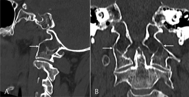
Figure 4
Sagittal (A) and coronal (B) reconstructions of a CT scan of the cervical spine shows assimilation of the massa lateralis of C1 and the occipital condyle (white arrows).