
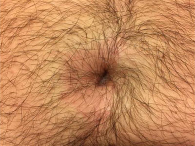
Figure 1
Periumbilical erythema.
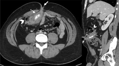
Figure 2
Coronal reformatted and axial contrast medium-enhanced CT-scan images showing dilated and thickened intestinal structure (dotted arrow) between the caecum (arrowhead) and the umbilicus (arrow), containing a calcified deposit.