
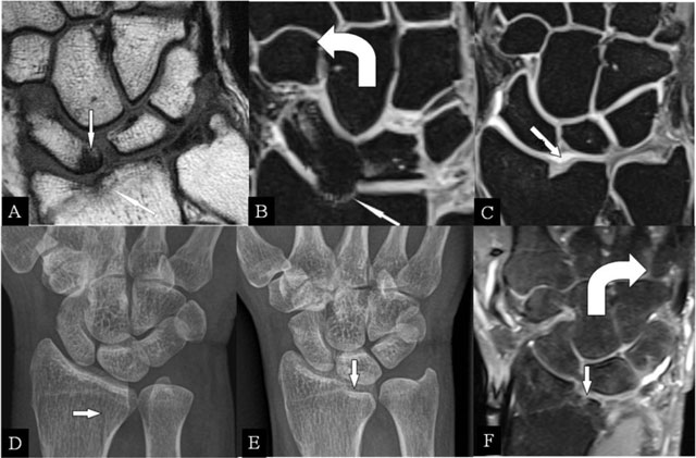
Figure 1
Radioscafoid and radiolunate abutment. (A) Coronal SE T1-WI; (B, C) Coronal 3D-GRE; (D, E) PA plain radiographs; and (F) Coronal SE PD-WI FS. (A) Sequela of an intra-articular fracture of the distal radial epiphysis with a residual step-off (oblique arrow) and marrow oedema (vertical arrow) at the proximal pole of the scaphoid bone. (B) Centrally, the cartilage is destroyed and the radial deviation is blocked. (C) Cartilage step-off in another patient. (D) The parasagittal intra-articular fracture was initially missed. (E) Consolidation with a depressed part of the articular surface. (F) Radiolunate abutment with blocked ulnar deviation.
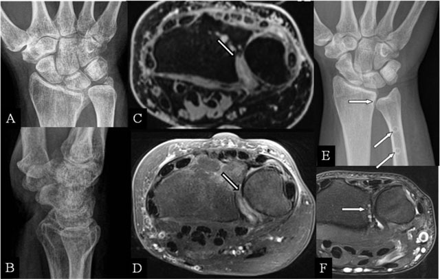
Figure 2
Radioulnar abutment and ulnar impingement. (A, B) PA and lateral plain radiographs; (C) Axial 3D-GRE; (D) SE T2-WI FS; (E) PA plain radiograph; and (F) Axial SE T2-WI FS. (A, B) Sequelae of a Pouteau-Colles fracture of the distal radial epiphysis. (C) Residual step-off at the radial sigmoid notch. (D) Destruction of the cartilage at the dorsal part of the sigmoid notch. (E) Excessive shortening (horizontal arrow) after surgery (oblique arrows). (F) subchondral erosions at the most proximal part of the radial sigmoid notch.
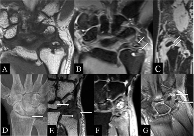
Figure 3
Ulnar (intra)styloid and stylotriquetral abutment. (A, E) Coronal SE T1-WI; (B, F) Coronal SE PD-WI FS; (C) Sagittal 3D-GRE; (D) PA plain radiograph; and (G) Coronal SE T1-WI FS with gadolinium. (A–C) Neoarticulation in the center of the ulnar styloid process, surrounding marrow oedema, (B) and juxta-articular cysts (arrows) (B, C). (D–F) Stylotriquetral abutment with flattening of the tip of the styloid process (D), bone marrow oedema and synovitis (E, F), and contrast enhancement of the marrow oedema and the synovitis (G).
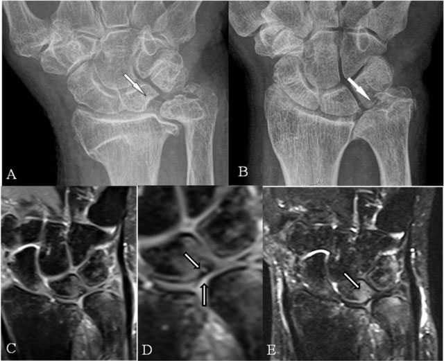
Figure 4
Ulnolunate and/or ulnotriquetral abutment. (A, B) PA plain radiographs; (C, D) Coronal SE PD-WI FS; and (E) Coronal SE T2-WI FS. (A) Ulnolunate abutment with a sclerotic defined impression at the ulnar side of the lunate bone. (B) Ulnotriquetral abutment with sclerotic bordered neoarticulation. (C–E) Ulnolunate abutment with chondromalacia at the ulnar border of the lunate bone (D, vertical arrow), subchondral cyst (D, oblique arrow), and bone marrow oedema centered at the ulnar side of the lunate bone (E).
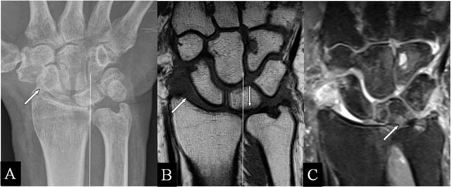
Figure 5
Ulnar translation with abutment. (A) PA plain radiograph; (B) Coronal SE T1-WI; and (C) Coronal SE PD-WI FS. (A–C) Lateral widening of the radioscaphoid joint (oblique arrow) and the lunate bone overlapping less than 50% with the corresponding radial articular fossa due to the ulnar translation of the carpus. (B, C) Cartilage destruction, oedema, and an accompanying lesion of the TFCC.
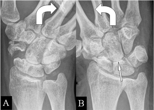
Figure 6
(Intra)scaphoid abutment. (A, B) PA plain mobility radiographs in ulnar and radial deviation. (A) Large diastasis in ulnar deviation between the scaphoid bone fragments. (B) Impaction of both fragments in radial deviation. Associated midcarpal osteoarthritis (arrow).
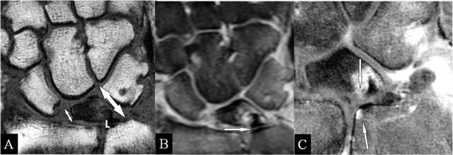
Figure 7
Lunar abutment. (A) Coronal SE T1-WI; (B) Coronal SE PD-WI FS; and (C) Coronal SE T1-WI FS with gadolinium. (A) Due to loss of height at the radial side, the ulnar side of the lunate bone (L) approaches the ulnar head. (B) Sclerotic borders at the contact zones (arrow), oedema at the ulnar corner of the lunate bone and TFCC tear. (C) Contrast enhancement in the zones of the kissing marrow oedema.
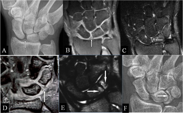
Figure 8
Scapholunar and lunotriquetral abutment. (A) PA plain radiograph; (B) Coronal SE PD-WI FS; (C) Coronal SE T2-WI FS; (D) Coronal 2D-GRE; (E) Coronal SE T2-WI FS; and (F) PA plain radiograph. (A) Widening of the scapholunate joint space on a Schneck I view. (B) Tear of the scapholunate ligament. (C) Juxta-articular subchondral band shaped marrow oedema. (D) Tear of the lunotriquetral ligament. (E) Juxta-articular band shaped kissing marrow oedema. (F) Massive deformation at the triquetral bone (other patient).
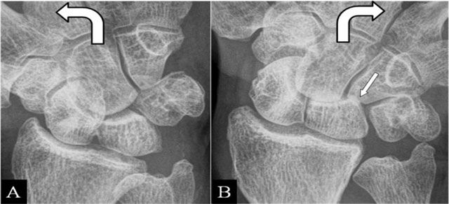
Figure 9
Hamatolunar abutment. (A) PA plain mobility radiographs in radial and ulnar deviation. (A, B) Step-off at the lunotriquetral joint of the first and second line of Gilula. (B) Secondary impaction of the hamate and lunate bone in ulnar deviation (oblique arrow).
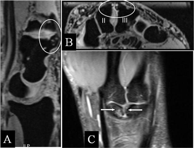
Figure 10
Carpal boss with abutment. (A) Sagittal 3D-GRE; (B) Coronal 3D-GRE; and (C) Coronal SE PD-WI FS. (A, B) Old posttraumatic deformation with subchondral cysts at the dorsum of the capitate bone (A) and the base of the second and third metacarpal bone (B) with misalignment around the quadrangular joint. (C) Juxta-articular kissing bone marrow oedema.