
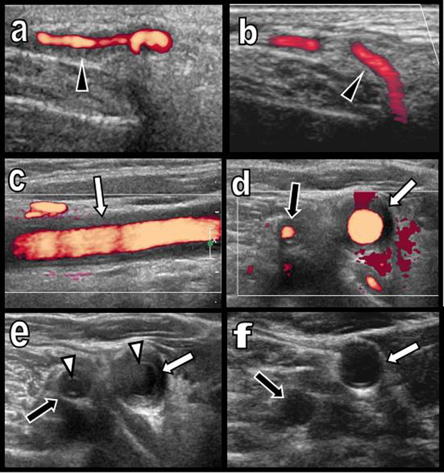
Figure 1
Colour Doppler Ultrasound of the temporal artery before (a) and after treatment (b) and of the right primitive carotid and vertebral arteries before (c, d, e) and after treatment (f).
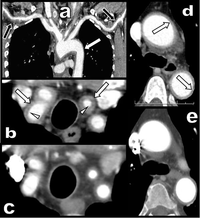
Figure 2
CT angiography of the aortic arch and of its large emerging arteries before (a, b, d) and after treatment (c and e).

Figure 3
Histopathology of the temporal artery biopsy.