
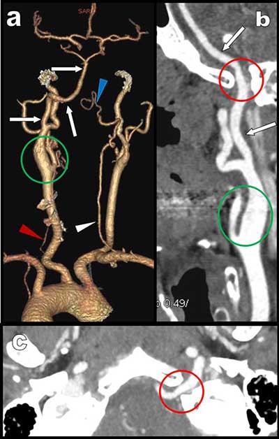
Figure 1
CTA: a) posterior-anterior VR view show major hypoplasia of the vertebral arteries. They are not connected to the basilar trunk which appear only feed by a large ascending artery emerging from a large left internal carotid. Curvilinear reconstruction (b) and axial MIP view (c) at the level of the foramen magna show this large hypoglossal artery penetrating the skull through the supracondylar hypoglossus canal.
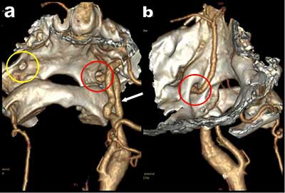
Figure 2
3D volume rendering views with bone structures showing the hypoglossal artery penetrating the skull through the supracondylar hypoglossal fossa.
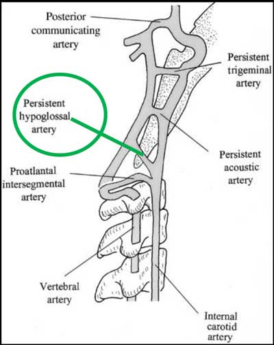
Figure 3
Schematic representation of the different fetal anastomoses between the vertebral arterial system and the carotid arterial system.