
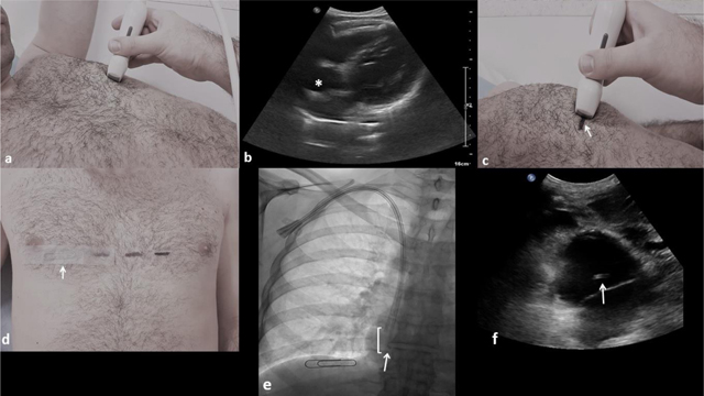
Figure 1
(a) The US probe is placed transversely in a left anterior intercostal space. (b) The right atrium is localized (asterisk) by a left intercostal approach. (c) A horizontal line is drawn on the skin (arrow) at the probe’s level (mid-width). (d) A paper clip (arrow) is stick on the skin at right (midclavicular line), at the same horizontal level that the drawn skin marker, corresponding to the atrial floor. (e) Antero-posterior fluoroscopic image of the chest showing a 23 cm inserted long term hemodialysis catheter through the right internal jugular vein with its distal tip (arrow) located above the paper clip, so the entire functional part (square bracket) is located inside the right atrium. (f) Subxiphoid US view confirming the distal catheter tip position inside the right atrium (arrow).