
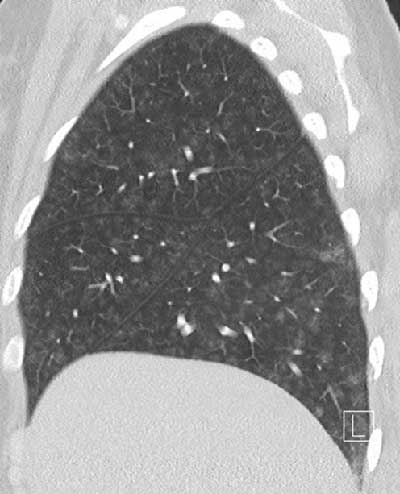
Figure 1
Diffuse groundglass centrilobular nodules without tree-in-bud.
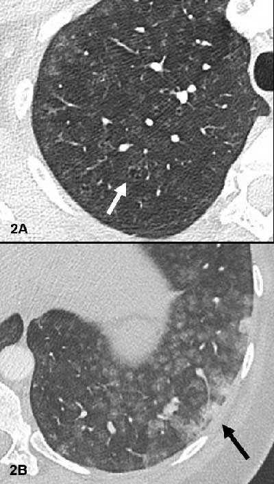
Figure 2
Small centrilobular apical emphysema (white arrow Figure 2A) and confluent condensed areas in the basal segments of the lower lobes (black arrow Figure 2B).
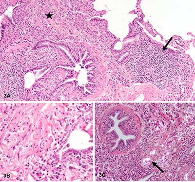
Figure 3
Important non-caseating granulomatous interstitial inflammation, with lymphocytes (black arrow Figure 3A), numerous macrophages and multinuclear giant cells, sitting preferentially in peribronchiolar regions (black star Figure 3A, B). The granulomas contain characteristic needle-shaped birefringent crystalline material in polarized light (black arrow Figure 3C).