
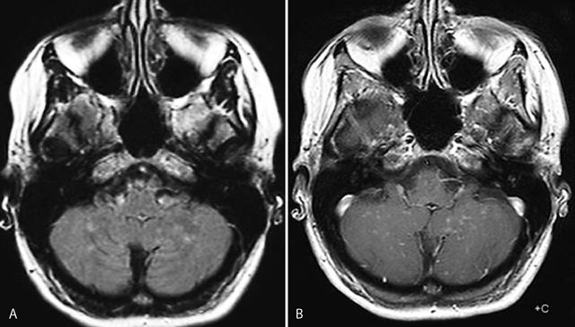
Figure 1
Axial FLAIR image (A): multiple punctate and linear FLAIR hyperintense blurry lesions without mass effect (A) predominatly located in the cerebellar vermis and hemispheres, which become more apperent on contrast-enhanced T1-weighted image (B).

Figure 2
Axial T1 with contrast showing multiple discrete curvilinear lesions in the cerebral white matter of the superior frontal and prerolandic gyrus in both hemispheres.
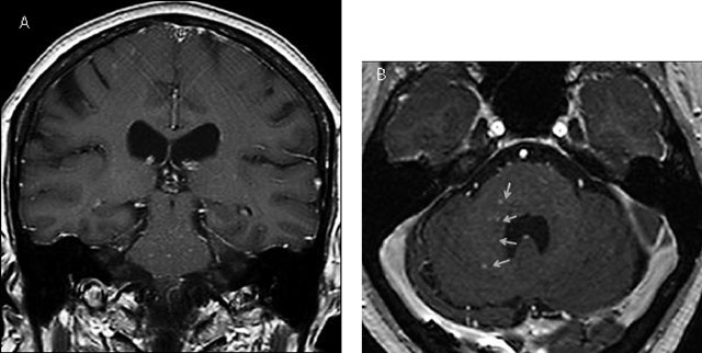
Figure 3
Coronal (A) and axial (B) contrast-enhanced T1 MRI images after 1.5 years showing new punctate and curvilinear lesions predomanintly in the pons and the right middle cerebellar peduncle (yellow arrows).