
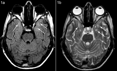
Figure 1
Patient A, axial FLAIR (a) and T2-weighted (b) images at the same level show subcortical white matter hyperintensity anteromedial in the temporal lobes, more pronounced on the left side.
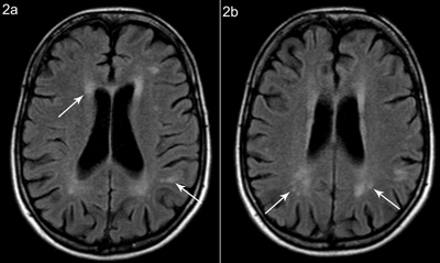
Figure 2
Patient A, axial FLAIR weighted images at different sections show (a) periventricular white matter hyperintensity and multiple hyperintense lesions in the subcortical white matter, with partial confluence and (b) white matter hyperintensity posterior and superior to the trigone.

Figure 3
Patient A, axial T2-weighted image shows prominent Virchow-Robin spaces bilateral in the centrum semiovale.
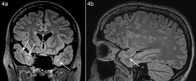
Figure 4
Patient B, coronal (a) and sagittal (b) FLAIR image shows white matter hyperintensity subcortical anteromedial in the temporal lobe.