
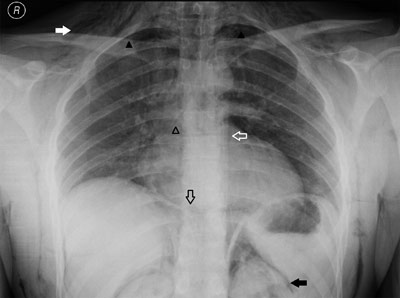
Figure A
Anteroposterior (AP) chest radiograph demonstrating diffuse subcutaneous emphysema (solid white arrow), a small pneumothorax in both apical lung fields (solid black arrowheads) and air outlining the thoracic aorta (hollow white arrow), the bronchial branches (hollow black arrowhead), both sides of the diaphragm (hollow black arrow) and both kidneys (solid black arrow).
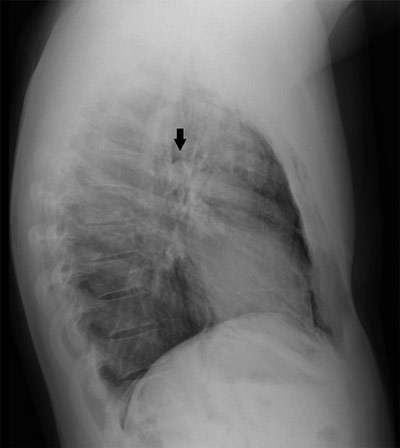
Figure B
Lateral chest radiograph showing a well-defined lucency surrounding the right pulmonary artery, also known as the ring around the artery sign (solid black arrow).
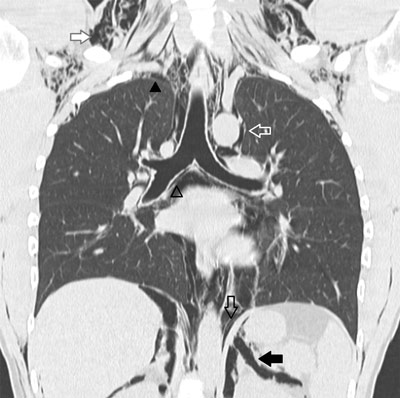
Figure C
CT of the chest confirming a pneumomediastinum (hollow black arrowhead, hollow black arrow and hollow white arrow) associated with diffuse subcutaneous emphysema (solid white arrow), pneumo(retro)peritoneum (solid black arrow) and pneumothorax (solid black arrowhead).