
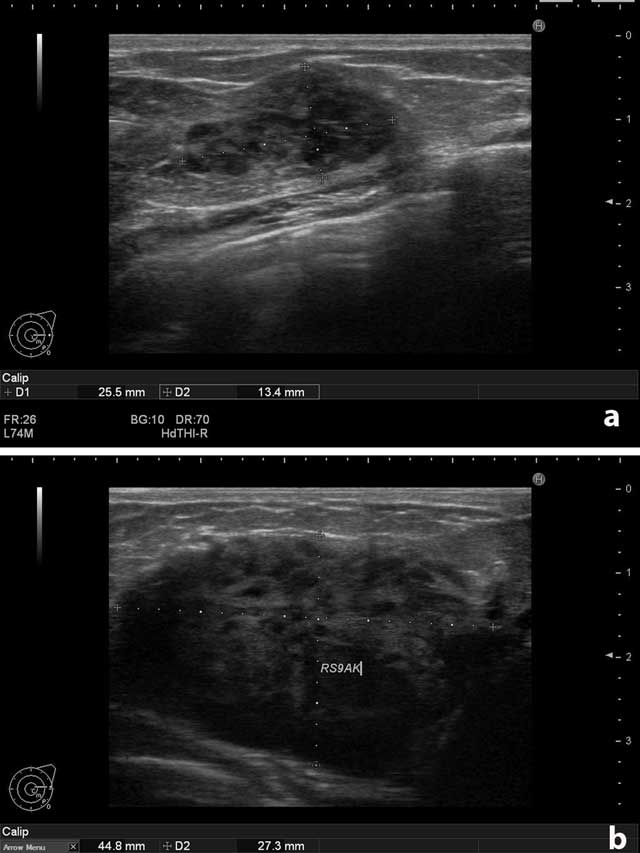
Figure 1
(a) Left breast. Solid, circumscribed mass. (b) Right breast. The largest solid mass measured 4.5 × 2.7 cm.
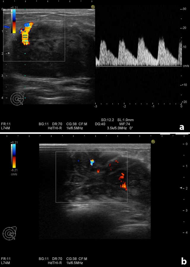
Figure 2
Doppler examination of the largest mass on the right breast. (a) Increased vascularity at the contours of the mass. (b) Increased vascularity in the internal part of the mass.
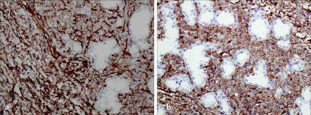
Figure 3
Histopathologic examination. (a) Strong positivity for CD34, ×200 (Figure 3a) and (b) CD43, ×200.