
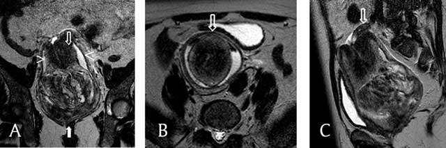
Figure 1
T2-weighted MRI (a, b, c) a. This coronal image shows a vaginal heterogeneous mass (filled arrow), with the uterine corpus in a U-shape above the mass (empty arrow). The cervix surrounds the corpus, and the vaginal fornix surrounds both the corpus and the cervix (arrowheads). b. This axial image shows, from the center outwards, the uterine corpus, the cervix, and the fornix and the invaginated round ligaments in a bullseye appearance (arrow). c. This sagittal image shows one ovary above the cervix (arrow).
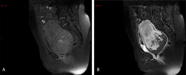
Figure 2
Sagittal T1-weighted (a, b) a. The bright spots of suppressed fat in the image denote hemorrhagic areas. b. After an injection of gadolinium, there is a heterogeneous enhancement of the mass.
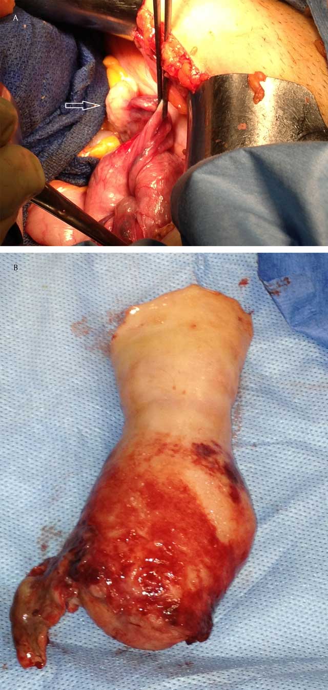
Figure 3
This operative view during a laparotomy shows the fallopian tube and one of the ovaries (arrow) outside the inverted uterus (a) and a completely reversed uterus after resection (b).