
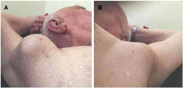
Figure 1
External appearance of the bilateral axillary pulsatile masses. (A) right, (B) Left.
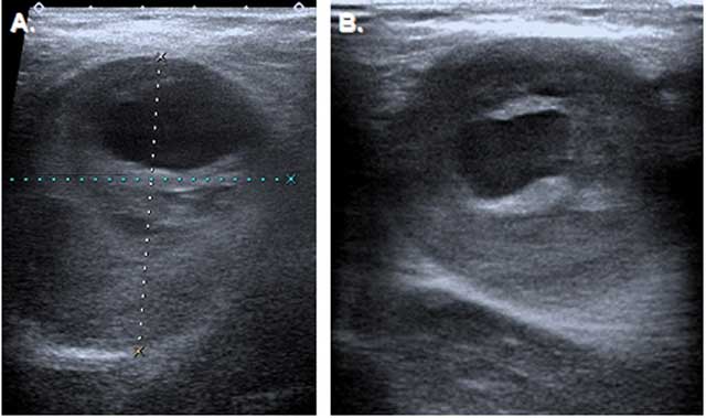
Figure 2
Ultrasonography of the axillary masses. (A) Transverse section show 67 × 45 mm partially thrombosed aneurysm on the right, (B) 40 × 45 mm partially thrombosed aneurysm on the left.
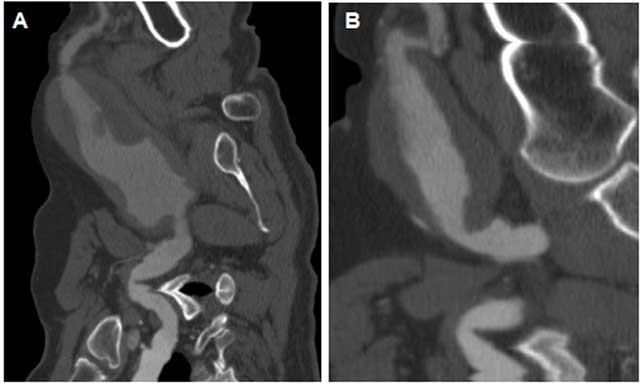
Figure 3
On a CT angiogram, bilateral axillary artery aneurysms were detected on curvilinear reformatted sections. (A) right, (B) Left.