
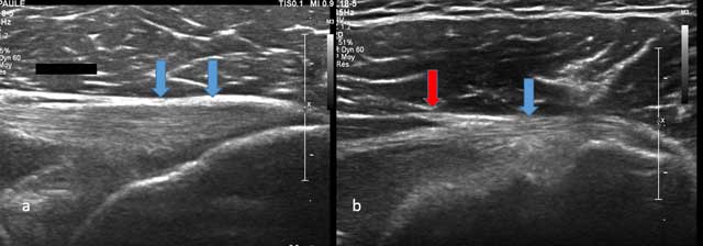
Figure 1
Axial view of Teres Minor with tendon (blue arrow) and musculotendinous junction (red arrow).
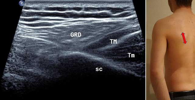
Figure 2
Sagittal oblique view of the scapula (Sc) in external position (red arrow). GRD: Latissimus Dorsi, TM: Teres Major, Tm: Teres minor.
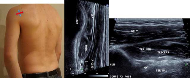
Figure 3
Sagittal oblique view (blue arrow) and axial intermediate view (red arrow) with HUM: Humerus, ISP: Infraspinatus, TMIN: Teres minor, A CIRC: Axillar Artery, DELT: Deltoid Muscle, TER MAJ: Teres Major, QV: Velpeau space, Triceps: Tricipal Muscle.
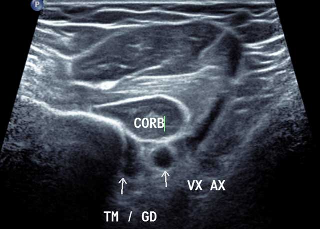
Figure 4
Axial distal section of the anterior and medial humerus area in external rotation. CORB: Coraco Brachal Muscle, TM/GD: Common enthesis of Teres major and Latissimus Dorsi, VX AX: Axillar Artery.
Figure 5
Sagittal view of the humeral diaphysis.
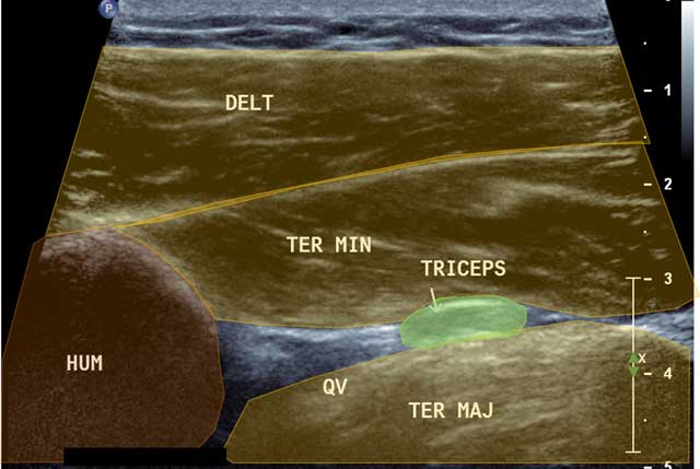
Figure 6
Focus in axial view of the quadrilateral space. HUM: Humerus, DELT: Deltoid muscle, QV: Quadrilateral Velpeau Space, Ter Maj: Teres Major, Ter Min: Teres Minor, Triceps: Tricipital muscle.
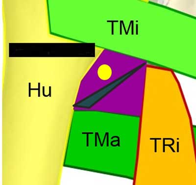
Figure 7
Coronal view of the QVadrilateral space. TMi: Teres Minor, TMa: Teres Major, TRi: Tricipital muscle, Yellow circle: Axillar nerve.
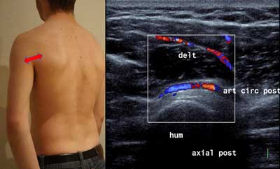
Figure 8
Axial posterior view of the humerus. delt: Deltoid muscle, hum: humerus, art circ post: Axillar artery.
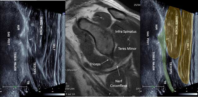
Figure 9
Sagittal view of the triceps tendon with MRI correlation. HUM: Humerus, TR: Triceps muscle, Nerf circonflexe: circumflex nerve.
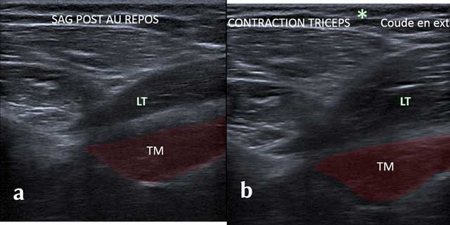
Figure 10
Sagittal view without (a) and with (b) Tricepscontraction with elbow in extension. TM: Teres Major, LT: long head of the triceps.
Figure 11
Sagittal view of the rotatorcuff with “coma” appearance of the myotendinous junction of the supra spinatus.
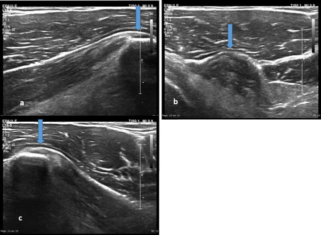
Figure 12
Axial view of the coracoid process with enthesis of the short head of the biceps brachii (a and b) and the coracobrachialis (c).
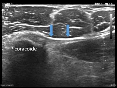
Figure 13
Axial view of the medial part of the coracoid process with enthesis of the pectoralis minor (arrows).
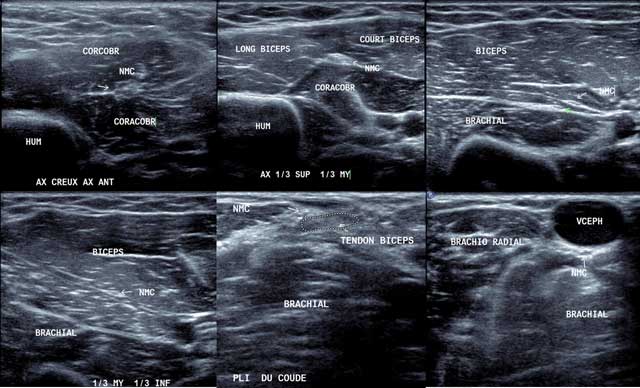
Figure 14
Axial view of the musculocutaneous nerve from the axillar area to the elbow (a to f). NMC: Musculocutaneous nerve, Coracobr: coracobrachial muscle, Hum: Humerus, Long Biceps: Long head of the biceps, Court biceps: short head of the biceps, Brachial: Brachial muscle, Brachio-radial: Brachio-radial muscle, VCEPH: Cephalic vein.
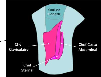
Figure 15
Pectoralis Major tendon on the humerus. Chef Sternal: sternal muscle, Chef Claviculaire: Clavicular muscle, Chef Costo Abdominal: Costo-abdominal muscle, Coulisse Bicipitale: Bicipital area.
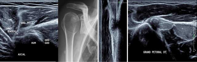
Figure 16
Axial view of the normal anterior part of the humerus in external position (a) with hydroxyapatite deposits in X-rays (b) and US in sagittal and axial view (d). GRD/GDO: Teres Major/Latissimus Dorsi, GRD PECT: Pectoralis Major, HUM: Humerus, Court Biceps: Short head of the biceps.
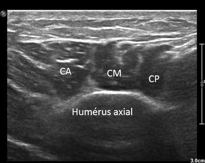
Figure 17
Axial view of the distal and lateral part of the deltoid muscle. CA: Anterior chief, CM: Medium chief, CP: Posterior chief.
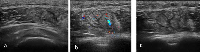
Figure 18
Axial view of a recent traumatic extrinsic lesion of the medium chief of the deltoid with hyperhemia (a–c).