
Figure 1
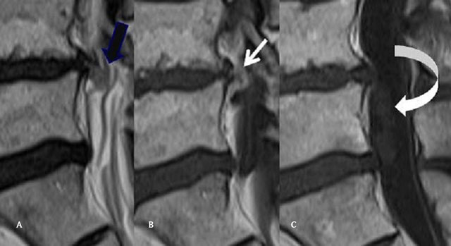
(A) Sagittal T2 Mri of the lumbar spine: Continuity of the disc and the upper part of the disc herniation (back arrow). (B) Sagittal T1 Mri of the lumbar spine, after gadolinium injection. The upper part of the disc is compact with peripheral contrast enhancement (white arrow). (C) Sagittal T1 Mri of the lumbar spine, after gadolinium injection. The intradural component of the IDH does not enhance (curved arrow).
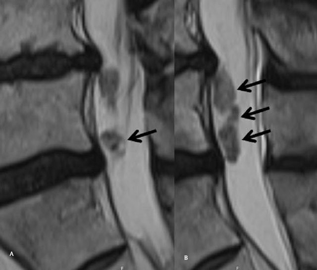
Figure 2A,B
Sagittal T2 Mri of the lumbar spine, “crumbled” aspect of the intradural component of the IDH (arrows).
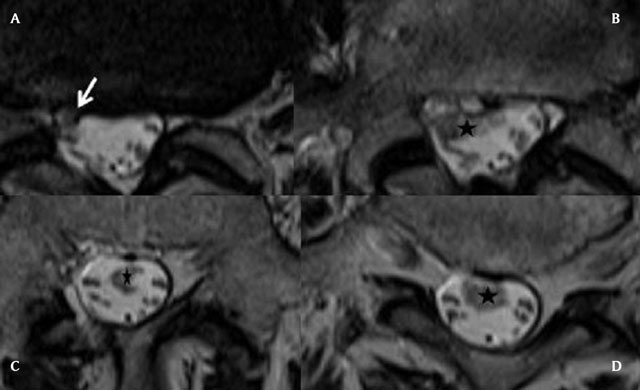
Figure 3A–D
Axial T2 slices of the lumbar spine, from the level of the L4-L5 disk to the sacrum. The IDH enters at the L4-L5 level (white arrow) and extends downwards in the CSF, to the L5-S1 level (black stars). It has irregular borders, heterogeneous nodular structure and variable diameter.
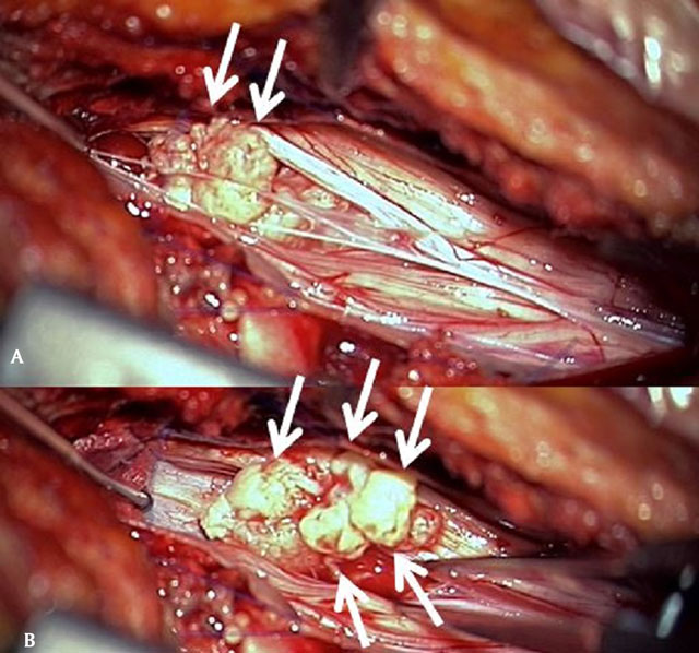
Figure 4A,B
Peroperative views after parasagittal opening of the dura. Multiple nodular fragments of a spongious disc herniation are extracted.