
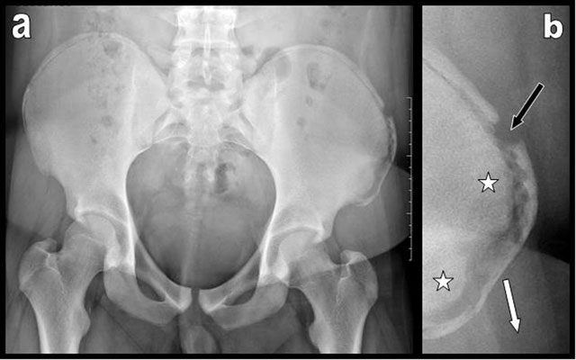
Figure 1
Pelvic plain film (a) and focused view (b) of the left iliac crest illustrate avulsion of the anterior part of the left iliac crest apophysis. Diastasis between the parent bone and the apophysis is clearly visible. The apophysis is displaced outwardly and downwardly (white arrow). Transverse fracture of the iliac crest apophysis itself is associated (black arrow). Preexisting bony sclerosis of the parent iliac bone due to chronic overuse is visible (white stars).
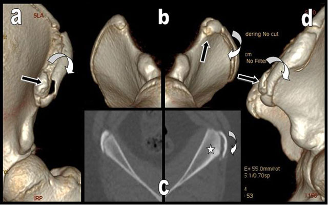
Figure 2
Left anterior (a), comparative right and left oblique inferior (b) and left lateral (d) 3D CT reconstructions of the left iliac crests illustrate the avulsion of the anterior part of the left iliac crest apophysis. It is displaced outwardly and downwardly (curved white arrow) but the apophysis of the anterior and superior iliac spine - origin of the sartorius - is respected and not displaced (black arrow). Comparative axial views of the right and left iliac crests (c) show preexisting hypertrophy and bony sclerosis of the left parent iliac bone due to chronic overuse (white star).
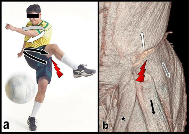
Figure 3
Typical posture of a right-handed soccer during fast kicking (a) volume rendering CT view of the muscles inserting on the iliac crest of our patient (b). Kicking is initiated by rotating the pelvis far to the left (curved black arrow on a) around the supporting left leg and by bringing forwards the thigh of the right kicking leg while maintaining the upper trunk to the right (curved white arrow on a). The sudden traction and contraction of the left external oblique (white arrow) probably pulls the apophysis off the left iliac crest. The action of the external oblique muscle is abruptly thwarted (red flash) by the antagonist traction of the gluteus medius (grey arrow) but merely of the tensor fascia lata (black arrow) that displaces the avulsed apophysis outwardly and downwardly. Black star = Sartorius.
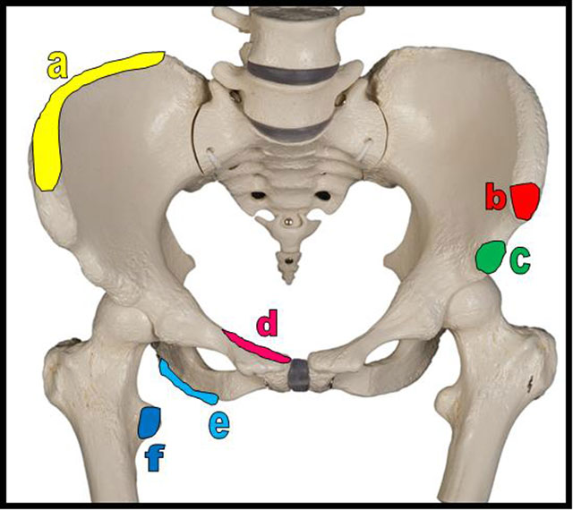
Figure 4
Schema of the most frequent sites of pelvic apophyseal avulsion fractures. a = iliac crest (insertions of the abdominal muscles, the tensor of fascia lata and of the gluteus medius); b = anterior superior iliac spine (insertion of sartorius); c = anterior inferior iliac spine (insertion of rectus femoris); d = superior corner of pubic symphysis (insertion of rectus abdominis); e = ischial tuberosity (insertion of hamstrings muscles = semitendinous, semimembranous and long head of the biceps femoris muscles); f = lesser trochanter (insertion of iliopsoas muscle).