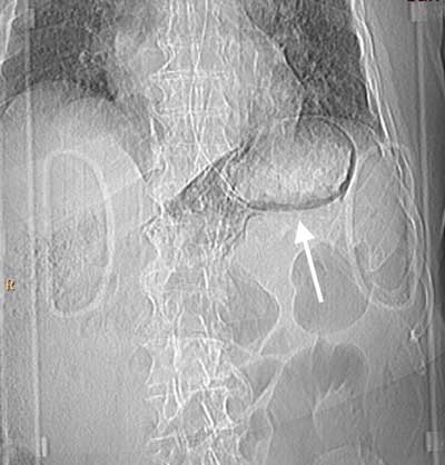
Have a personal or library account? Click to login
Gastric Emphysema after Intubation Cover

Figures & Tables

jbsr-101-1-812-g1.jpg
Figure 1

Topogram preliminary to the CT scan. Linear gas shadows in the upper left abdominal region (arrow), apparently round the stomach.
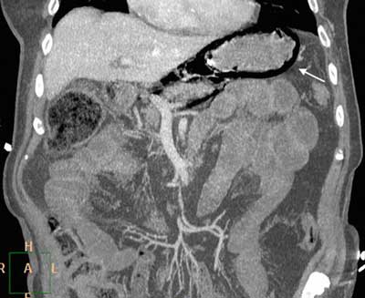
jbsr-101-1-812-g2.jpg
Figure 2

Contrast-enhanced CT, MPR reformats. Large amounts of intramural gas in the stomach (arrow) with no evidence of free peritoneal air.
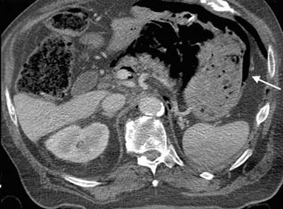
jbsr-101-1-812-g3.jpg
Figure 3

Contrast-enhanced CT, axial slice. Evidence of gastric intramural gas (arrow) and air bubbles within the retroperitoneal space.

DOI: https://doi.org/10.5334/jbr-btr.812 | Journal eISSN: 2514-8281
Language: English
Published on: Jan 10, 2017
Published by: Ubiquity Press
In partnership with: Paradigm Publishing Services
Publication frequency: 1 issue per year

© 2017 Valeria Angelino, Giovanni Volpicelli, Luciano Cardinale, published by Ubiquity Press
This work is licensed under the Creative Commons Attribution 4.0 License.