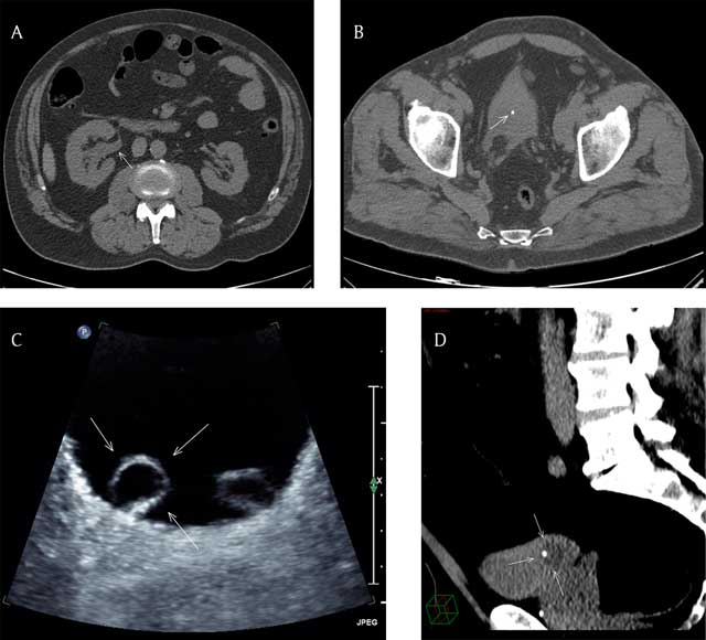
Have a personal or library account? Click to login
Obstructive Ureterocele Stone Mimicking an Intravesical Stone Cover

Obstructive Ureterocele Stone Mimicking an Intravesical Stone

By: Sammy Tawk and  Etienne Danse  
Open Access
|Jan 2018

Figures & Tables

jbsr-101-1-1433-g1.jpg
Figure 1

(A) Axial CT scan image (non-contrast) at the level of the abdomen showing right uretero-pyelo-calyceal dilatation (arrow). (B) Axial CT scan image (non-contrast) at the level of the pelvis showing a 3.6 mm hyperdense stone in an almost empty bladder (arrow). (C) Axial US image of the bladder showing bilateral ureterocele, more prominent at right (arrows). (D) Reformatted sagittal CT scan (non-contrast) image with narrow window width of the pelvis showing the urine filled right ureterocele with the stone located inside (arrows).

Language: English
Published on: Jan 3, 2018
Published by: Ubiquity Press
In partnership with: Paradigm Publishing Services
Publication frequency: 1 issue per year

© 2018 Sammy Tawk, Etienne Danse, published by Ubiquity Press
This work is licensed under the Creative Commons Attribution 4.0 License.